- PDB-4jpq: Crystal structure of a putative carbohydrate-binding protein (BAC... -
+
Open data
ID or keywords:
Loading...
-
Basic information
Entry
Database: PDB / ID: 4jpq
Title
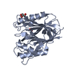
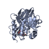
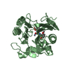
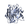
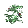
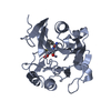
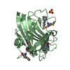
Crystal structure of a putative carbohydrate-binding protein (BACUNI_03838) from Bacteroides uniformis ATCC 8492 at 2.70 A resolution
Components
Uncharacterized protein
Keywords
STRUCTURAL GENOMICS / UNKNOWN FUNCTION / Carbohydrate-binding family 9 / PF16011 / Joint Center for Structural Genomics / JCSG / Protein Structure Initiative / PSI-BIOLOGY
Function / homology
Function and homology information
carbohydrate catabolic process / hydrolase activity, hydrolyzing O-glycosyl compounds / carbohydrate binding Similarity search - Function
Mass: 18.015 Da / Num. of mol.: 29 / Source method: isolated from a natural source / Formula: H2O
Has protein modification
Y
Sequence details
THIS CONSTRUCT WAS EXPRESSED WITH A PURIFICATION TAG MGSDKIHHHHHHENLYFQG. THE TAG WAS REMOVED WITH ...THIS CONSTRUCT WAS EXPRESSED WITH A PURIFICATION TAG MGSDKIHHHHHHENLYFQG. THE TAG WAS REMOVED WITH TEV PROTEASE LEAVING ONLY A GLYCINE (0) FOLLOWED BY RESIDUES 32-250 OF THE TARGET SEQUENCE.
-
Experimental details
-
Experiment
Experiment
Method: X-RAY DIFFRACTION / Number of used crystals: 1
-
Sample preparation
Crystal
Density Matthews: 3.26 Å3/Da / Density % sol: 62.27 %
Crystal grow
Temperature: 277 K / Method: vapor diffusion, sitting drop / pH: 10.5 Details: 0.2000M lithium sulfate, 2.0000M ammonium sulfate, 0.1M CAPS pH 10.5, NANODROP, VAPOR DIFFUSION, SITTING DROP, temperature 277K
Type: DECTRIS PILATUS 6M / Detector: PIXEL / Date: Jan 23, 2013 Details: Flat mirror (vertical focusing); single crystal Si(111) bent monochromator (horizontal focusing)
Radiation
Monochromator: single crystal Si(111) bent / Protocol: MAD / Monochromatic (M) / Laue (L): M / Scattering type: x-ray
Radiation wavelength
ID
Wavelength (Å)
Relative weight
1
0.91837
1
2
0.97941
1
3
0.97858
1
Reflection
Resolution: 2.7→29.23 Å / Num. obs: 18547 / % possible obs: 99.2 % / Observed criterion σ(I): -3 / Biso Wilson estimate: 73.497 Å2 / Rmerge(I) obs: 0.095 / Net I/σ(I): 13.68
Reflection shell
Rmerge(I) obs: 0.01 / Diffraction-ID: 1
Resolution (Å)
Highest resolution (Å)
Mean I/σ(I) obs
Num. measured obs
Num. unique obs
% possible all
2.7-2.8
1.8
14802
3460
97.2
2.8-2.91
2.7
14218
3328
99.5
2.91-3.04
3.3
13073
3354
98.8
3.04-3.2
5.5
15030
3413
99.5
3.2-3.4
8.8
15422
3418
99.8
3.4-3.66
11.7
15190
3416
99.9
3.66-4.03
17
15227
3479
99.9
4.03-4.6
22.9
13316
3357
99.3
4.6-5.77
27.2
15445
3423
99.9
5.77
35.3
14971
3499
98.4
-
Phasing
Phasing
Method: MAD
-
Processing
Software
Name
Version
Classification
NB
MolProbity
3beta29
modelbuilding
PDB_EXTRACT
3.1
dataextraction
SHELX
phasing
SHARP
phasing
XSCALE
July4, 2012
datascaling
REFMAC
5.7.0032
refinement
XDS
datareduction
SHELXD
phasing
Refinement
Method to determine structure: MAD / Resolution: 2.7→29.23 Å / Cor.coef. Fo:Fc: 0.945 / Cor.coef. Fo:Fc free: 0.927 / Occupancy max: 1 / Occupancy min: 0.5 / SU B: 25.357 / SU ML: 0.236 / Cross valid method: THROUGHOUT / σ(F): 0 / ESU R: 0.58 / ESU R Free: 0.295 Stereochemistry target values: MAXIMUM LIKELIHOOD WITH PHASES Details: 1. HYDROGENS HAVE BEEN ADDED IN THE RIDING POSITIONS. 2. ATOM RECORD CONTAINS SUM OF TLS AND RESIDUAL B FACTORS. 3. ANISOU RECORD CONTAINS SUM OF TLS AND RESIDUAL U FACTORS. 4. WATERS WERE ...Details: 1. HYDROGENS HAVE BEEN ADDED IN THE RIDING POSITIONS. 2. ATOM RECORD CONTAINS SUM OF TLS AND RESIDUAL B FACTORS. 3. ANISOU RECORD CONTAINS SUM OF TLS AND RESIDUAL U FACTORS. 4. WATERS WERE EXCLUDED FROM AUTOMATIC TLS ASSIGNMENT. 5. A MET-INHIBITION PROTOCOL WAS USED FOR SELENOMETHIONINE INCORPORATION DURING PROTEIN EXPRESSION. THE OCCUPANCY OF THE SE ATOMS IN THE MSE RESIDUES WAS REDUCED TO 0.75 FOR THE REDUCED SCATTERING POWER DUE TO PARTIAL S-MET INCORPORATION. 6. SULFATE ION (SO4) AND 3-CYCLOHEXYL-1-PROPYLSULFONIC ACID (CXS) FROM THE CRYSTALLIZATION SOLUTION ARE MODELED.
Rfactor
Num. reflection
% reflection
Selection details
Rfree
0.2381
946
5.1 %
RANDOM
Rwork
0.2056
-
-
-
obs
0.2071
18497
99.29 %
-
Solvent computation
Ion probe radii: 0.8 Å / Shrinkage radii: 0.8 Å / VDW probe radii: 1.2 Å / Solvent model: BABINET MODEL WITH MASK
In the structure databanks used in Yorodumi, some data are registered as the other names, "COVID-19 virus" and "2019-nCoV". Here are the details of the virus and the list of structure data.
Jan 31, 2019. EMDB accession codes are about to change! (news from PDBe EMDB page)
EMDB accession codes are about to change! (news from PDBe EMDB page)
The allocation of 4 digits for EMDB accession codes will soon come to an end. Whilst these codes will remain in use, new EMDB accession codes will include an additional digit and will expand incrementally as the available range of codes is exhausted. The current 4-digit format prefixed with “EMD-” (i.e. EMD-XXXX) will advance to a 5-digit format (i.e. EMD-XXXXX), and so on. It is currently estimated that the 4-digit codes will be depleted around Spring 2019, at which point the 5-digit format will come into force.
The EM Navigator/Yorodumi systems omit the EMD- prefix.
Related info.:Q: What is EMD? / ID/Accession-code notation in Yorodumi/EM Navigator
Yorodumi is a browser for structure data from EMDB, PDB, SASBDB, etc.
This page is also the successor to EM Navigator detail page, and also detail information page/front-end page for Omokage search.
The word "yorodu" (or yorozu) is an old Japanese word meaning "ten thousand". "mi" (miru) is to see.
Related info.:EMDB / PDB / SASBDB / Comparison of 3 databanks / Yorodumi Search / Aug 31, 2016. New EM Navigator & Yorodumi / Yorodumi Papers / Jmol/JSmol / Function and homology information / Changes in new EM Navigator and Yorodumi
 Movie
Movie Controller

 Yorodumi
 Open data
 Basic information
 Components
 Keywords
 Function and homology information
 Bacteroides uniformis (bacteria)
 X-RAY DIFFRACTION /
  Authors
 Citation
 Structure visualization
 Downloads & links
 Other downloads


 PDBj
PDBj Assembly
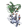


 Mass: 221.317 Da / Num. of mol.: 3 / Source method: obtained synthetically / Formula: C9H19NO3S / Comment: pH buffer*YM
 Mass: 96.063 Da / Num. of mol.: 6 / Source method: obtained synthetically / Formula: SO4
 Mass: 18.015 Da / Num. of mol.: 29 / Source method: isolated from a natural source / Formula: H2O
 Sample preparation
 / Beamline: BL11-1 / Wavelength: 0.91837,0.97941,0.97858
 Processing