 Movie
Movie Controller


+ Open data

- Basic information
| Entry | Database: PDB / ID: 4le4 | |||||||||
|---|---|---|---|---|---|---|---|---|---|---|
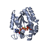
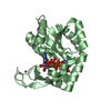
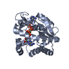
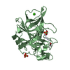
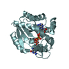
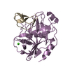
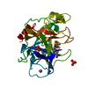
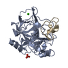
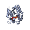
| Title | Crystal structure of PaGluc131A with cellotriose | |||||||||
 Components Components | Beta-glucanase | |||||||||
 Keywords Keywords | HYDROLASE / glucanse / GH131 | |||||||||
| Function / homology |  Function and homology information Function and homology informationcellulose binding / carbohydrate metabolic process / extracellular region Similarity search - Function | |||||||||
| Biological species |  Podospora anserina (fungus) Podospora anserina (fungus) | |||||||||
| Method |  X-RAY DIFFRACTION / SYNCHROTRON / MOLECULAR REPLACEMENT / Resolution: 1.8 Å X-RAY DIFFRACTION / SYNCHROTRON / MOLECULAR REPLACEMENT / Resolution: 1.8 Å | |||||||||
 Authors Authors | Jiang, T. / Chan, H.C. / Huang, C.H. / Ko, T.P. / Huang, T.Y. / Liu, J.R. / Guo, R.T. | |||||||||
 Citation Citation | Journal: To be Published Title: Crystal Structures of a GH131 beta-Glucanase Catalytic Domain from Podospora anserina in Complex with Cellotriose Authors: Jiang, T. / Chan, H.C. / Huang, C.H. / Ko, T.P. / Huang, T.Y. / Liu, J.R. / Guo, R.T. | |||||||||
| History |
|
- Structure visualization
| Structure viewer | Molecule: MolmilJmol/JSmol |
|---|
- Downloads & links
-Download
| PDBx/mmCIF format | 4le4.cif.gz | 225.8 KB | Display | PDBx/mmCIF format |
|---|---|---|---|---|
| PDB format | pdb4le4.ent.gz | 179.4 KB | Display | PDB format |
| PDBx/mmJSON format | 4le4.json.gz | Tree view | PDBx/mmJSON format | |
| Others |  Other downloads Other downloads |
-Validation report
| Arichive directory | https://data.pdbj.org/pub/pdb/validation_reports/le/4le4ftp://data.pdbj.org/pub/pdb/validation_reports/le/4le4 | HTTPS FTP |
|---|
-Related structure data
| Related structure data | 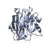 4le3S S: Starting model for refinement |
|---|---|
| Similar structure data |

-Links
 PDBj
PDBj- Assembly
| Deposited unit | 
| ||||||||
|---|---|---|---|---|---|---|---|---|---|
| 1 | 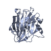
| ||||||||
| 2 | 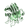
| ||||||||
| 3 | 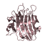
| ||||||||
| 4 | 
| ||||||||
| Unit cell |
|
-Components
| #1: Protein | Mass: 27718.361 Da / Num. of mol.: 4 / Fragment: UNP residues 19-270 Source method: isolated from a genetically manipulated source Source: (gene. exp.) Podospora anserina (fungus) / Plasmid: pET32 Xa/LIC / Production host:  #2: Polysaccharide | beta-D-glucopyranose-(1-4)-beta-D-glucopyranose-(1-4)-beta-D-glucopyranose / beta-cellotriose |   Source method: isolated from a genetically manipulated source Details: oligosaccharide / References: beta-cellotriose #3: Water | ChemComp-HOH / |  Mass: 18.015 Da / Num. of mol.: 1116 / Source method: isolated from a natural source / Formula: H2O Mass: 18.015 Da / Num. of mol.: 1116 / Source method: isolated from a natural source / Formula: H2O |
|---|
-Experimental details
-Experiment
| Experiment | Method: X-RAY DIFFRACTION / Number of used crystals: 1 |
|---|
- Sample preparation
| Crystal | Density Matthews: 2.26 Å3/Da / Density % sol: 45.59 % |
|---|---|
| Crystal grow | Temperature: 298 K / Method: vapor diffusion, sitting drop / pH: 7 Details: 0.1M HEPES, 0.7M LiCl, 33% PEG6K, pH 7.0, VAPOR DIFFUSION, SITTING DROP, temperature 298K |
-Data collection
| Diffraction | Mean temperature: 100 K |
|---|---|
| Diffraction source | Source: SYNCHROTRON / Site: NSRRC  / Beamline: BL13B1 / Wavelength: 0.9762 Å / Beamline: BL13B1 / Wavelength: 0.9762 Å |
| Detector | Type: ADSC QUANTUM 315r / Detector: CCD / Date: May 3, 2013 |
| Radiation | Protocol: SINGLE WAVELENGTH / Monochromatic (M) / Laue (L): M / Scattering type: x-ray |
| Radiation wavelength | Wavelength: 0.9762 Å / Relative weight: 1 |
| Reflection | Resolution: 1.8→25 Å / Num. obs: 87110 / % possible obs: 97 % / Redundancy: 3.4 % |
| Reflection shell | Resolution: 1.8→1.86 Å / Redundancy: 3.3 % / Rmerge(I) obs: 0.297 / Mean I/σ(I) obs: 3 / Num. unique all: 8638 / % possible all: 95.9 |
- Processing
| Software |
| ||||||||||||||||||||
|---|---|---|---|---|---|---|---|---|---|---|---|---|---|---|---|---|---|---|---|---|---|
| Refinement | Method to determine structure: MOLECULAR REPLACEMENT Starting model: 4LE3 Resolution: 1.8→25 Å / σ(F): 3 / Stereochemistry target values: Engh & Huber
| ||||||||||||||||||||
| Refinement step | Cycle: LAST / Resolution: 1.8→25 Å
| ||||||||||||||||||||
| LS refinement shell | Resolution: 1.8→1.86 Å /
|
