 Movie
Movie Controller

[English] 日本語
 Yorodumi
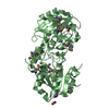
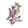
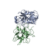
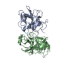
- PDB-1avw: COMPLEX PORCINE PANCREATIC TRYPSIN/SOYBEAN TRYPSIN INHIBITOR, ORT... -
+ Open data

- Basic information
| Entry | Database: PDB / ID: 1avw | ||||||
|---|---|---|---|---|---|---|---|
| Title | COMPLEX PORCINE PANCREATIC TRYPSIN/SOYBEAN TRYPSIN INHIBITOR, ORTHORHOMBIC CRYSTAL FORM | ||||||
 Components Components |
| ||||||
 Keywords Keywords | COMPLEX (PROTEINASE/INHIBITOR) / COMPLEX (PROTEINASE-INHIBITOR) / PORCINE TRYPSIN / SOYBEAN TRYPSIN INHIBITOR / KUNITZ-TYPE / BETA-TREFOIL FOLD / COMPLEX (PROTEINASE-INHIBITOR) complex | ||||||
| Function / homology |  Function and homology information Function and homology informationtrypsin / digestion / serine-type endopeptidase inhibitor activity / serine-type endopeptidase activity / proteolysis / : / metal ion binding Similarity search - Function | ||||||
| Biological species |   | ||||||
| Method |  X-RAY DIFFRACTION / SYNCHROTRON / MOLECULAR REPLACEMENT / Resolution: 1.75 Å X-RAY DIFFRACTION / SYNCHROTRON / MOLECULAR REPLACEMENT / Resolution: 1.75 Å | ||||||
 Authors Authors | Song, H.K. / Suh, S.W. | ||||||
 Citation Citation | Journal: J.Mol.Biol. / Year: 1998 Title: Kunitz-type soybean trypsin inhibitor revisited: refined structure of its complex with porcine trypsin reveals an insight into the interaction between a homologous inhibitor from Erythrina ...Title: Kunitz-type soybean trypsin inhibitor revisited: refined structure of its complex with porcine trypsin reveals an insight into the interaction between a homologous inhibitor from Erythrina caffra and tissue-type plasminogen activator. Authors: Song, H.K. / Suh, S.W. #1: Journal: Thesis, Seoul National University / Year: 1997Title: Crystal Structure Analyses of Human A1-Antitrypsin, Soybean Kunitz-Type Trypsin Inhibitor, and Barley Chitinase Authors: Song, H.K. #2: Journal: Mol.Cells / Year: 1993Title: Crystallization of Kunitz-Type Soybean Trypsin Inhibitor Authors: Lee, J.K. / Song, H.K. / Hwang, K.Y. / Kim, K.K. / Suh, S.W. #3: Journal: Thesis, Seoul National University / Year: 1993Title: Crystallization and Preliminary X-Ray Crystallographic Study of Kunitz-Type Soybean Trypsin Inhibitor Authors: Lee, J.K. | ||||||
| History |
|
- Structure visualization
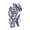
| Structure viewer | Molecule: MolmilJmol/JSmol |
|---|
- Downloads & links
-Download
| PDBx/mmCIF format | 1avw.cif.gz | 89.6 KB | Display | PDBx/mmCIF format |
|---|---|---|---|---|
| PDB format | pdb1avw.ent.gz | 66.6 KB | Display | PDB format |
| PDBx/mmJSON format | 1avw.json.gz | Tree view | PDBx/mmJSON format | |
| Others |  Other downloads Other downloads |
-Validation report
| Arichive directory | https://data.pdbj.org/pub/pdb/validation_reports/av/1avwftp://data.pdbj.org/pub/pdb/validation_reports/av/1avw | HTTPS FTP |
|---|
-Related structure data
| Related structure data |  1avuC  1avxC 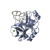 1mctS  1tieS S: Starting model for refinement C: citing same article ( |
|---|---|
| Similar structure data |


-Links
 PDBj
PDBj

- Assembly
| Deposited unit | 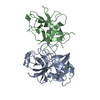
| ||||||||
|---|---|---|---|---|---|---|---|---|---|
| 1 |
| ||||||||
| Unit cell |
|
-Components
| #1: Protein | Mass: 23493.496 Da / Num. of mol.: 1 / Source method: isolated from a natural source / Source: (natural) |
|---|---|
| #2: Protein | Mass: 19657.109 Da / Num. of mol.: 1 / Source method: isolated from a natural source / Source: (natural) |
| #3: Chemical | ChemComp-CA /   Mass: 40.078 Da / Num. of mol.: 1 / Source method: obtained synthetically / Formula: Ca Mass: 40.078 Da / Num. of mol.: 1 / Source method: obtained synthetically / Formula: Ca |
| #4: Water | ChemComp-HOH /  Mass: 18.015 Da / Num. of mol.: 142 / Source method: isolated from a natural source / Formula: H2O Mass: 18.015 Da / Num. of mol.: 142 / Source method: isolated from a natural source / Formula: H2O |
| Has protein modification | Y |
-Experimental details
-Experiment
| Experiment | Method: X-RAY DIFFRACTION / Number of used crystals: 1 |
|---|
- Sample preparation
| Crystal | Density Matthews: 3.16 Å3/Da / Density % sol: 61 % | ||||||||||||||||||||
|---|---|---|---|---|---|---|---|---|---|---|---|---|---|---|---|---|---|---|---|---|---|
| Crystal grow | pH: 7.5 / Details: pH 7.5 | ||||||||||||||||||||
| Crystal | *PLUS Density % sol: 61 % | ||||||||||||||||||||
| Crystal grow | *PLUS pH: 7.67 / Method: vapor diffusion, hanging drop | ||||||||||||||||||||
| Components of the solutions | *PLUS
|
-Data collection
| Diffraction | Mean temperature: 293 K |
|---|---|
| Diffraction source | Source: SYNCHROTRON / Site: NSLS  / Beamline: X12C / Wavelength: 1 / Beamline: X12C / Wavelength: 1 |
| Detector | Type: MARRESEARCH / Detector: IMAGE PLATE AREA DETECTOR / Date: Sep 1, 1994 |
| Radiation | Monochromatic (M) / Laue (L): M / Scattering type: x-ray |
| Radiation wavelength | Wavelength: 1 Å / Relative weight: 1 |
| Reflection | Resolution: 1.75→50 Å / Num. obs: 52687 / % possible obs: 92 % / Observed criterion σ(I): 0.5 / Redundancy: 7.8 % / Rmerge(I) obs: 0.078 |
| Reflection shell | Resolution: 1.75→1.8 Å / % possible all: 68.6 |
| Reflection | *PLUS Num. measured all: 419433 |
| Reflection shell | *PLUS % possible obs: 68.6 % |
- Processing
| Software |
| ||||||||||||||||||||||||||||||||||||||||||||||||||||||||||||
|---|---|---|---|---|---|---|---|---|---|---|---|---|---|---|---|---|---|---|---|---|---|---|---|---|---|---|---|---|---|---|---|---|---|---|---|---|---|---|---|---|---|---|---|---|---|---|---|---|---|---|---|---|---|---|---|---|---|---|---|---|---|
| Refinement | Method to determine structure: MOLECULAR REPLACEMENT Starting model: PDB ENTRY 1MCT FOR TRYPSIN AND PDB ENTRY 1TIE FOR THE INHIBITOR Resolution: 1.75→8 Å / Data cutoff high absF: 0 / Data cutoff low absF: 0 / σ(F): 2
| ||||||||||||||||||||||||||||||||||||||||||||||||||||||||||||
| Refine analyze | Luzzati d res low obs: 0 Å | ||||||||||||||||||||||||||||||||||||||||||||||||||||||||||||
| Refinement step | Cycle: LAST / Resolution: 1.75→8 Å
| ||||||||||||||||||||||||||||||||||||||||||||||||||||||||||||
| Refine LS restraints |
| ||||||||||||||||||||||||||||||||||||||||||||||||||||||||||||
| Xplor file |
|
