 Movie
Movie Controller

[English] 日本語
 Yorodumi
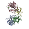
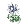
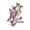
- PDB-3i29: Crystal structure of a binary complex between an mutant trypsin i... -
+ Open data

- Basic information
| Entry | Database: PDB / ID: 3i29 | ||||||
|---|---|---|---|---|---|---|---|
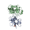
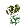
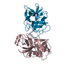
| Title | Crystal structure of a binary complex between an mutant trypsin inhibitor with bovine trypsin | ||||||
 Components Components |
| ||||||
 Keywords Keywords | HYDROLASE/HYDROLASE INHIBITOR / P1 and P2 MUTANT / TRYPSIN INHIBITOR / BOVINE TRYPSIN / COMPLEX / DIGESTION / HYDROLASE / METAL-BINDING / PROTEASE / SECRETED / SERINE PROTEASE / ZYMOGEN / PROTEASE INHIBITOR / SERINE PROTEASE INHIBITOR / HYDROLASE-HYDROLASE INHIBITOR COMPLEX / Disulfide bond | ||||||
| Function / homology |  Function and homology information Function and homology informationtrypsin / serpin family protein binding / serine protease inhibitor complex / digestion / serine-type endopeptidase inhibitor activity / endopeptidase activity / serine-type endopeptidase activity / proteolysis / : / metal ion binding Similarity search - Function | ||||||
| Biological species |    Psophocarpus tetragonolobus (winged bean) Psophocarpus tetragonolobus (winged bean) | ||||||
| Method |  X-RAY DIFFRACTION / MOLECULAR REPLACEMENT / Resolution: 2.4 Å X-RAY DIFFRACTION / MOLECULAR REPLACEMENT / Resolution: 2.4 Å | ||||||
 Authors Authors | Khamrui, S. / Majumder, S. / Dasgupta, J. / Dattagupta, J.K. / Sen, U. | ||||||
 Citation Citation | Journal: Biochim.Biophys.Acta / Year: 2012 Title: Role of remote scaffolding residues in the inhibitory loop pre-organization, flexibility, rigidification and enzyme inhibition of serine protease inhibitors Authors: Majumder, S. / Khamrui, S. / Dasgupta, J. / Dattagupta, J.K. / Sen, U. | ||||||
| History |
|
- Structure visualization
| Structure viewer | Molecule: MolmilJmol/JSmol |
|---|
- Downloads & links
-Download
| PDBx/mmCIF format | 3i29.cif.gz | 92 KB | Display | PDBx/mmCIF format |
|---|---|---|---|---|
| PDB format | pdb3i29.ent.gz | 68.1 KB | Display | PDB format |
| PDBx/mmJSON format | 3i29.json.gz | Tree view | PDBx/mmJSON format | |
| Others |  Other downloads Other downloads |
-Validation report
| Arichive directory | https://data.pdbj.org/pub/pdb/validation_reports/i2/3i29ftp://data.pdbj.org/pub/pdb/validation_reports/i2/3i29 | HTTPS FTP |
|---|
-Related structure data
| Related structure data |  3qydC  3veqC 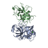 1avwS  1eylS S: Starting model for refinement C: citing same article ( |
|---|---|
| Similar structure data |


-Links
 PDBj
PDBj

- Assembly
| Deposited unit | 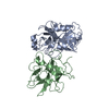
| ||||||||
|---|---|---|---|---|---|---|---|---|---|
| 1 |
| ||||||||
| Unit cell |
|
-Components
| #1: Protein | Mass: 23324.287 Da / Num. of mol.: 1 Source method: isolated from a genetically manipulated source Source: (gene. exp.)  |
|---|---|
| #2: Protein | Mass: 20740.357 Da / Num. of mol.: 1 / Mutation: F64Y, L65R Source method: isolated from a genetically manipulated source Source: (gene. exp.) Psophocarpus tetragonolobus (winged bean)Plasmid: pET28a+ / Production host: |
| #3: Chemical | ChemComp-CA /   Mass: 40.078 Da / Num. of mol.: 1 / Source method: obtained synthetically / Formula: Ca Mass: 40.078 Da / Num. of mol.: 1 / Source method: obtained synthetically / Formula: Ca |
| #4: Water | ChemComp-HOH /  Mass: 18.015 Da / Num. of mol.: 125 / Source method: isolated from a natural source / Formula: H2O Mass: 18.015 Da / Num. of mol.: 125 / Source method: isolated from a natural source / Formula: H2O |
| Has protein modification | Y |
-Experimental details
-Experiment
| Experiment | Method: X-RAY DIFFRACTION / Number of used crystals: 1 |
|---|
- Sample preparation
| Crystal | Density Matthews: 1.84 Å3/Da / Density % sol: 33.18 % |
|---|---|
| Crystal grow | Temperature: 298 K / Method: vapor diffusion, hanging drop / pH: 8 Details: pH 8.0, VAPOR DIFFUSION, HANGING DROP, temperature 298K |
-Data collection
| Diffraction source | Source: ROTATING ANODE / Type: BRUKER AXS MICROSTAR / Wavelength: 1.54 Å |
|---|---|
| Detector | Type: MAR scanner 345 mm plate / Detector: IMAGE PLATE / Date: Aug 8, 2008 |
| Radiation | Protocol: SINGLE WAVELENGTH / Monochromatic (M) / Laue (L): M / Scattering type: x-ray |
| Radiation wavelength | Wavelength: 1.54 Å / Relative weight: 1 |
| Reflection | Resolution: 2.4→30 Å / Num. obs: 12051 / Redundancy: 2.87 % / Biso Wilson estimate: 29.9 Å2 / Rmerge(I) obs: 0.1073 / Net I/σ(I): 4.2 |
- Processing
| Software | Name: CNS / Version: 1.2 / Classification: refinement | ||||||||||||||||||||||||||||||||||||||||||||||||||||||||||||
|---|---|---|---|---|---|---|---|---|---|---|---|---|---|---|---|---|---|---|---|---|---|---|---|---|---|---|---|---|---|---|---|---|---|---|---|---|---|---|---|---|---|---|---|---|---|---|---|---|---|---|---|---|---|---|---|---|---|---|---|---|---|
| Refinement | Method to determine structure: MOLECULAR REPLACEMENT Starting model: PDB ENTRIES 1AVW (for trypsin) AND 1EYL (for inhibitor) Resolution: 2.4→9.99 Å / Rfactor Rfree error: 0.012 / Data cutoff high absF: 1284624.5 / Data cutoff low absF: 0 / Isotropic thermal model: RESTRAINED / Cross valid method: THROUGHOUT / σ(F): 0 / Details: BULK SOLVENT MODEL USED
| ||||||||||||||||||||||||||||||||||||||||||||||||||||||||||||
| Solvent computation | Solvent model: FLAT MODEL / Bsol: 64.826 Å2 / ksol: 0.5 e/Å3 | ||||||||||||||||||||||||||||||||||||||||||||||||||||||||||||
| Displacement parameters | Biso mean: 30.3 Å2
| ||||||||||||||||||||||||||||||||||||||||||||||||||||||||||||
| Refine analyze |
| ||||||||||||||||||||||||||||||||||||||||||||||||||||||||||||
| Refinement step | Cycle: LAST / Resolution: 2.4→9.99 Å
| ||||||||||||||||||||||||||||||||||||||||||||||||||||||||||||
| Refine LS restraints |
| ||||||||||||||||||||||||||||||||||||||||||||||||||||||||||||
| LS refinement shell | Resolution: 2.4→2.55 Å / Rfactor Rfree error: 0.035 / Total num. of bins used: 6
| ||||||||||||||||||||||||||||||||||||||||||||||||||||||||||||
| Xplor file |
|
