 Movie
Movie Controller

[English] 日本語
 Yorodumi
- PDB-1osb: Conjugative Relaxase TrwC in complex with OriT Dna. Metal-free st... -
+ Open data

- Basic information
| Entry | Database: PDB / ID: 1osb | ||||||
|---|---|---|---|---|---|---|---|
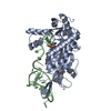
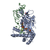
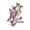
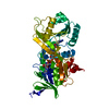
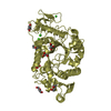
| Title | Conjugative Relaxase TrwC in complex with OriT Dna. Metal-free structure. | ||||||
 Components Components |
| ||||||
 Keywords Keywords | TRANSFERASE/DNA / Bacterial conjugation / Relaxase / DNA replication / TRANSFERASE-DNA COMPLEX | ||||||
| Function / homology | Conjugative relaxase, N-terminal / TrwC relaxase / TrwC relaxase / AAA domain / P-loop containing nucleoside triphosphate hydrolase / metal ion binding / DNA / DNA (> 10) / TrwC Function and homology information Function and homology information | ||||||
| Biological species |  | ||||||
| Method |  X-RAY DIFFRACTION / SYNCHROTRON / MOLECULAR REPLACEMENT / Resolution: 2.65 Å X-RAY DIFFRACTION / SYNCHROTRON / MOLECULAR REPLACEMENT / Resolution: 2.65 Å | ||||||
 Authors Authors | Guasch, A. / Lucas, M. / Moncalian, G. / Cabezas, M. / Perez-Luque, R. / Gomis-Ruth, F.X. / de la Cruz, F. / Coll, M. | ||||||
 Citation Citation | Journal: Nat.Struct.Biol. / Year: 2003 Title: Recognition and processing of the origin of transfer DNA by conjugative relaxase TrwC. Authors: Guasch, A. / Lucas, M. / Moncalian, G. / Cabezas, M. / Perez-Luque, R. / Gomis-Ruth, F.X. / de la Cruz, F. / Coll, M. #1: Journal: J.Mol.Biol. / Year: 2000Title: Two active-site tyrosyl residues of protein TrwC act sequentially at the origin of transfer during plasmid R388 conjugation Authors: Grandoso, G. / Avila, P. / Cayon, A. / Hernando, M.A. / Llosa, M. / de la Cruz, F. #2: Journal: Mol.Microbiol. / Year: 2002Title: Bacterial conjugation: a two-step mechanism for DNA transport. Authors: Llosa, M. / Gomis-Ruth, F.X. / Coll, M. / de la Cruz, F. | ||||||
| History |
|
- Structure visualization
| Structure viewer | Molecule: MolmilJmol/JSmol |
|---|
- Downloads & links
-Download
| PDBx/mmCIF format | 1osb.cif.gz | 157.8 KB | Display | PDBx/mmCIF format |
|---|---|---|---|---|
| PDB format | pdb1osb.ent.gz | 121.5 KB | Display | PDB format |
| PDBx/mmJSON format | 1osb.json.gz | Tree view | PDBx/mmJSON format | |
| Others |  Other downloads Other downloads |
-Validation report
| Arichive directory | https://data.pdbj.org/pub/pdb/validation_reports/os/1osbftp://data.pdbj.org/pub/pdb/validation_reports/os/1osb | HTTPS FTP |
|---|
-Related structure data
| Related structure data |  1omhSC  1qx0C S: Starting model for refinement C: citing same article ( |
|---|---|
| Similar structure data |


-Links
 PDBj
PDBj


- Assembly
| Deposited unit | 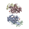
| ||||||||
|---|---|---|---|---|---|---|---|---|---|
| 1 | 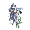
| ||||||||
| 2 | 
| ||||||||
| Unit cell |
|
-Components
| #1: DNA chain | Mass: 7714.970 Da / Num. of mol.: 2 / Source method: obtained synthetically / Details: DNA forming a cruciform arm #2: Protein | Mass: 32894.832 Da / Num. of mol.: 2 / Fragment: N-terminal relaxase domain Source method: isolated from a genetically manipulated source Source: (gene. exp.) #3: Chemical | ChemComp-SO4 /   Mass: 96.063 Da / Num. of mol.: 9 / Source method: obtained synthetically / Formula: SO4 Mass: 96.063 Da / Num. of mol.: 9 / Source method: obtained synthetically / Formula: SO4#4: Water | ChemComp-HOH / |  Mass: 18.015 Da / Num. of mol.: 257 / Source method: isolated from a natural source / Formula: H2O Mass: 18.015 Da / Num. of mol.: 257 / Source method: isolated from a natural source / Formula: H2O |
|---|
-Experimental details
-Experiment
| Experiment | Method: X-RAY DIFFRACTION / Number of used crystals: 1 |
|---|
- Sample preparation
| Crystal | Density Matthews: 3.03 Å3/Da / Density % sol: 59.41 % | ||||||||||||||||||||||||||||||
|---|---|---|---|---|---|---|---|---|---|---|---|---|---|---|---|---|---|---|---|---|---|---|---|---|---|---|---|---|---|---|---|
| Crystal grow | Temperature: 296 K / Method: vapor diffusion, hanging drop / pH: 4.6 Details: 25% PEGMM 2000, 0.2 M AMMONIUM SULPHATE, 0.1 M SODIUM ACETATE, pH 4.6, VAPOR DIFFUSION, HANGING DROP, temperature 296KK | ||||||||||||||||||||||||||||||
| Components of the solutions |
| ||||||||||||||||||||||||||||||
| Crystal grow | *PLUS Temperature: 4 ℃ | ||||||||||||||||||||||||||||||
| Components of the solutions | *PLUS
|
-Data collection
| Diffraction | Mean temperature: 100 K |
|---|---|
| Diffraction source | Source: SYNCHROTRON / Site: ESRF  / Beamline: ID13 / Beamline: ID13 |
| Detector | Type: MACSCIENCE / Detector: CCD / Date: Feb 20, 2002 |
| Radiation | Monochromator: diamond / Protocol: SINGLE WAVELENGTH / Monochromatic (M) / Laue (L): M / Scattering type: x-ray |
| Radiation wavelength | Relative weight: 1 |
| Reflection | Resolution: 2.65→40 Å / Num. obs: 28480 / % possible obs: 99.5 % / Observed criterion σ(F): 1 / Observed criterion σ(I): 1 / Redundancy: 4.4 % / Rmerge(I) obs: 0.098 / Net I/σ(I): 7 |
| Reflection shell | Highest resolution: 2.65 Å / Redundancy: 3.7 % / Rmerge(I) obs: 0.291 / Mean I/σ(I) obs: 2.5 / % possible all: 97.1 |
- Processing
| Software |
| |||||||||||||||||||||||||
|---|---|---|---|---|---|---|---|---|---|---|---|---|---|---|---|---|---|---|---|---|---|---|---|---|---|---|
| Refinement | Method to determine structure: MOLECULAR REPLACEMENT Starting model: pdb entry 1OMH Resolution: 2.65→40 Å / Cross valid method: FREE R-VALUE / σ(F): 1 / Stereochemistry target values: Engh & Huber
| |||||||||||||||||||||||||
| Refinement step | Cycle: LAST / Resolution: 2.65→40 Å
| |||||||||||||||||||||||||
| Refine LS restraints |
| |||||||||||||||||||||||||
| LS refinement shell | Resolution: 2.65→2.719 Å /
|
