[English] 日本語
Yorodumi
- PDB-3zy2: Crystal structure of POFUT1 in complex with GDP (High resolution ... -

+
Open data


ID or keywords:

Loading...

-
Basic information

Entry
Database: PDB / ID: 3zy2
TitleCrystal structure of POFUT1 in complex with GDP (High resolution dataset)
ComponentsPUTATIVE GDP-FUCOSE PROTEIN O-FUCOSYLTRANSFERASE 1
KeywordsTRANSFERASE / GLYCOSYLTRANSFERASE / GT-B / CATALYTIC MECHANISM / GT65
Function / homology
Function and homology information


peptide-O-fucosyltransferase / protein O-linked fucosylation / peptide-O-fucosyltransferase activity / fucosyltransferase activity / fucose metabolic process / protein O-linked glycosylation / Notch signaling pathway / endoplasmic reticulum
Similarity search - Function
GDP-fucose protein O-fucosyltransferase 1 / GDP-fucose protein O-fucosyltransferase / GDP-fucose protein O-fucosyltransferase / Rossmann fold - #11340 / Rossmann fold - #11350 / Rossmann fold / 3-Layer(aba) Sandwich / Alpha Beta
Similarity search - Domain/homology
GUANOSINE-5'-DIPHOSPHATE / : / GDP-fucose protein O-fucosyltransferase 1
Similarity search - Component
Biological speciesCAENORHABDITIS ELEGANS (invertebrata)
MethodX-RAY DIFFRACTION / SYNCHROTRON / SAD / Resolution: 1.54 Å
AuthorsLira-Navarrete, E. / Valero-Gonzalez, J. / Villanueva, R. / Martinez-Julvez, M. / Tejero, T. / Merino, P. / Panjikar, S. / Hurtado-Guerrero, R.
CitationJournal: Plos One / Year: 2011
Title: Structural Insights Into the Mechanism of Protein O-Fucosylation.
Authors: Lira-Navarrete, E. / Valero-Gonzalez, J. / Villanueva, R. / Martinez-Julvez, M. / Tejero, T. / Merino, P. / Panjikar, S. / Hurtado-Guerrero, R.
History
DepositionAug 17, 2011Deposition site: PDBE / Processing site: PDBE
Revision 1.0Sep 14, 2011Provider: repository / Type: Initial release
Revision 1.1Oct 12, 2011Group: Database references

-
Structure visualization

Structure viewerMolecule:
MolmilJmol/JSmol

Downloads & links

-
Assembly

Deposited unit
A: PUTATIVE GDP-FUCOSE PROTEIN O-FUCOSYLTRANSFERASE 1
hetero molecules


Theoretical massNumber of molelcules
Total (without water)41,5073
Polymers41,0091
Non-polymers4982
Water6,323351
1
A: PUTATIVE GDP-FUCOSE PROTEIN O-FUCOSYLTRANSFERASE 1
hetero molecules

A: PUTATIVE GDP-FUCOSE PROTEIN O-FUCOSYLTRANSFERASE 1
hetero molecules


Theoretical massNumber of molelcules
Total (without water)83,0146
Polymers82,0172
Non-polymers9964
Water362
TypeNameSymmetry operationNumber
identity operation1_555x,y,z1
crystal symmetry operation2_555-x,y,-z1
Buried area2620 Å2
ΔGint-18.4 kcal/mol
Surface area29130 Å2
MethodPISA
Unit cell
Length a, b, c (Å)145.018, 38.247, 68.196
Angle α, β, γ (deg.)90.00, 103.07, 90.00
Int Tables number5
Space group name H-MC121

-
Components

#1: Protein PUTATIVE GDP-FUCOSE PROTEIN O-FUCOSYLTRANSFERASE 1 / PEPTIDE-O-FUCOSYLTRANSFERASE 1 / O-FUCT-1 / POFUT1


Mass: 41008.746 Da / Num. of mol.: 1 / Fragment: RESIDUES 26-383
Source method: isolated from a genetically manipulated source
Source: (gene. exp.) CAENORHABDITIS ELEGANS (invertebrata) / Production host: 
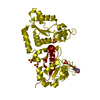
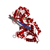
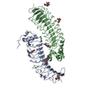
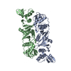
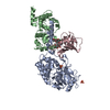
PICHIA PASTORIS (fungus) / Strain (production host): X-33 / References: UniProt: Q18014, peptide-O-fucosyltransferase
#2: Chemical ChemComp-MN / MANGANESE (II) ION


Mass: 54.938 Da / Num. of mol.: 1 / Source method: obtained synthetically / Formula: Mn
#3: Chemical ChemComp-GDP / GUANOSINE-5'-DIPHOSPHATE


Type: RNA linking / Mass: 443.201 Da / Num. of mol.: 1 / Source method: obtained synthetically / Formula: C10H15N5O11P2 / Comment: GDP, energy-carrying molecule*YM
#4: Water ChemComp-HOH / water


Mass: 18.015 Da / Num. of mol.: 351 / Source method: isolated from a natural source / Formula: H2O

-
Experimental details

-
Experiment

ExperimentMethod: X-RAY DIFFRACTION

-
Sample preparation

CrystalDensity Matthews: 2.23 Å3/Da / Density % sol: 44.86 %
Description: 3ZY2 WAS SOLVED FROM SAD PHASING METHODOLOGY USING A HIGHLY REDUNDANT DATASET TOGETHER WITH AN ISOMORPHOUS HIGH RESOLUTION DATASET (IN THIS CASE THIS ISOMORPHOUS HIGH RESOLUTION DATASET IS 3ZY2)

-
Data collection

DiffractionMean temperature: 90 K
Diffraction sourceSource: SYNCHROTRON / Site: ESRF / Beamline: BM16 / Wavelength: 0.99
DetectorType: MARRESEARCH / Detector: CCD
RadiationProtocol: SINGLE WAVELENGTH / Monochromatic (M) / Laue (L): M / Scattering type: x-ray
Radiation wavelengthWavelength: 0.99 Å / Relative weight: 1
ReflectionResolution: 1.54→19.5 Å / Num. obs: 53812 / % possible obs: 99.1 % / Observed criterion σ(I): 2 / Redundancy: 4.1 % / Rmerge(I) obs: 0.04 / Net I/σ(I): 20.8
Reflection shellResolution: 1.54→1.62 Å / Redundancy: 4 % / Rmerge(I) obs: 0.28 / Mean I/σ(I) obs: 4.8 / % possible all: 99.7

-
Processing

Software
NameVersionClassification
REFMAC5.5.0102refinement
XDSdata reduction
SCALEPACKdata scaling
RefinementMethod to determine structure: SAD
Starting model: NONE

Resolution: 1.54→70.63 Å / Cor.coef. Fo:Fc: 0.948 / Cor.coef. Fo:Fc free: 0.934 / SU B: 3.307 / SU ML: 0.057 / Cross valid method: THROUGHOUT / ESU R: 0.09 / ESU R Free: 0.09 / Stereochemistry target values: MAXIMUM LIKELIHOOD
Details: HYDROGENS HAVE BEEN ADDED IN THE RIDING POSITIONS. U VALUES WITH TLS ADDED.
RfactorNum. reflection% reflectionSelection details
Rfree0.23725 1010 1.9 %RANDOM
Rwork0.20507 ---
obs0.2057 52544 98.4 %-
Solvent computationIon probe radii: 0.8 Å / Shrinkage radii: 0.8 Å / VDW probe radii: 1.4 Å / Solvent model: MASK
Displacement parametersBiso mean: 23.687 Å2
Baniso -1Baniso -2Baniso -3
1--0.64 Å20 Å2-1.05 Å2
2--1.17 Å20 Å2
3----1 Å2
Refinement stepCycle: LAST / Resolution: 1.54→70.63 Å
ProteinNucleic acidLigandSolventTotal
Num. atoms2734 0 29 351 3114
Refine LS restraints
Refine-IDTypeDev idealDev ideal targetNumber
X-RAY DIFFRACTIONr_bond_refined_d0.0110.0222818
X-RAY DIFFRACTIONr_bond_other_d
X-RAY DIFFRACTIONr_angle_refined_deg1.3731.9453820
X-RAY DIFFRACTIONr_angle_other_deg
X-RAY DIFFRACTIONr_dihedral_angle_1_deg5.5935339
X-RAY DIFFRACTIONr_dihedral_angle_2_deg34.5523.817131
X-RAY DIFFRACTIONr_dihedral_angle_3_deg13.88215466
X-RAY DIFFRACTIONr_dihedral_angle_4_deg11.4071515
X-RAY DIFFRACTIONr_chiral_restr0.0910.2401
X-RAY DIFFRACTIONr_gen_planes_refined0.0070.0212191
X-RAY DIFFRACTIONr_gen_planes_other
X-RAY DIFFRACTIONr_nbd_refined
X-RAY DIFFRACTIONr_nbd_other
X-RAY DIFFRACTIONr_nbtor_refined
X-RAY DIFFRACTIONr_nbtor_other
X-RAY DIFFRACTIONr_xyhbond_nbd_refined
X-RAY DIFFRACTIONr_xyhbond_nbd_other
X-RAY DIFFRACTIONr_metal_ion_refined
X-RAY DIFFRACTIONr_metal_ion_other
X-RAY DIFFRACTIONr_symmetry_vdw_refined
X-RAY DIFFRACTIONr_symmetry_vdw_other
X-RAY DIFFRACTIONr_symmetry_hbond_refined
X-RAY DIFFRACTIONr_symmetry_hbond_other
X-RAY DIFFRACTIONr_symmetry_metal_ion_refined
X-RAY DIFFRACTIONr_symmetry_metal_ion_other
X-RAY DIFFRACTIONr_mcbond_it0.6541.51710
X-RAY DIFFRACTIONr_mcbond_other
X-RAY DIFFRACTIONr_mcangle_it1.20722774
X-RAY DIFFRACTIONr_mcangle_other
X-RAY DIFFRACTIONr_scbond_it2.17231108
X-RAY DIFFRACTIONr_scbond_other
X-RAY DIFFRACTIONr_scangle_it3.3244.51046
X-RAY DIFFRACTIONr_scangle_other
X-RAY DIFFRACTIONr_long_range_B_refined
X-RAY DIFFRACTIONr_long_range_B_other
X-RAY DIFFRACTIONr_rigid_bond_restr
X-RAY DIFFRACTIONr_sphericity_free
X-RAY DIFFRACTIONr_sphericity_bonded
LS refinement shellResolution: 1.54→1.58 Å / Total num. of bins used: 20
RfactorNum. reflection% reflection
Rfree0.391 75 -
Rwork0.27 3858 -
obs--99.12 %
Refinement TLS params.Method: refined / Origin x: 14.7158 Å / Origin y: -19.2162 Å / Origin z: 15.0536 Å
111213212223313233
T0.0561 Å20.0048 Å2-0.002 Å2-0.0081 Å20.0071 Å2--0.0628 Å2
L0.9405 °20.0792 °20.454 °2-0.1116 °20.0038 °2--1.919 °2
S0.0595 Å °0.0656 Å °-0.0245 Å °-0.0167 Å °0.0174 Å °0.0269 Å °0.0071 Å °-0.0288 Å °-0.0769 Å °

+
About Yorodumi

-
News

-
Feb 9, 2022. New format data for meta-information of EMDB entries

New format data for meta-information of EMDB entries

  • Version 3 of the EMDB header file is now the official format.
  • The previous official version 1.9 will be removed from the archive.

Related info.:EMDB header

External links:wwPDB to switch to version 3 of the EMDB data model

-
Aug 12, 2020. Covid-19 info

Covid-19 info

URL: https://pdbjlvh1.pdbj.org/emnavi/covid19.php

New page: Covid-19 featured information page in EM Navigator.

Related info.:Covid-19 info / Mar 5, 2020. Novel coronavirus structure data

+
Mar 5, 2020. Novel coronavirus structure data

Novel coronavirus structure data

Related info.:Yorodumi Speices / Aug 12, 2020. Covid-19 info

External links:COVID-19 featured content - PDBj / Molecule of the Month (242):Coronavirus Proteases

+
Jan 31, 2019. EMDB accession codes are about to change! (news from PDBe EMDB page)

EMDB accession codes are about to change! (news from PDBe EMDB page)

  • The allocation of 4 digits for EMDB accession codes will soon come to an end. Whilst these codes will remain in use, new EMDB accession codes will include an additional digit and will expand incrementally as the available range of codes is exhausted. The current 4-digit format prefixed with “EMD-” (i.e. EMD-XXXX) will advance to a 5-digit format (i.e. EMD-XXXXX), and so on. It is currently estimated that the 4-digit codes will be depleted around Spring 2019, at which point the 5-digit format will come into force.
  • The EM Navigator/Yorodumi systems omit the EMD- prefix.

Related info.:Q: What is EMD? / ID/Accession-code notation in Yorodumi/EM Navigator

External links:EMDB Accession Codes are Changing Soon! / Contact to PDBj

+
Jul 12, 2017. Major update of PDB

Major update of PDB

  • wwPDB released updated PDB data conforming to the new PDBx/mmCIF dictionary.
  • This is a major update changing the version number from 4 to 5, and with Remediation, in which all the entries are updated.
  • In this update, many items about electron microscopy experimental information are reorganized (e.g. em_software).
  • Now, EM Navigator and Yorodumi are based on the updated data.

External links:wwPDB Remediation / Enriched Model Files Conforming to OneDep Data Standards Now Available in the PDB FTP Archive

-
Yorodumi

Thousand views of thousand structures

  • Yorodumi is a browser for structure data from EMDB, PDB, SASBDB, etc.
  • This page is also the successor to EM Navigator detail page, and also detail information page/front-end page for Omokage search.
  • The word "yorodu" (or yorozu) is an old Japanese word meaning "ten thousand". "mi" (miru) is to see.

Related info.:EMDB / PDB / SASBDB / Comparison of 3 databanks / Yorodumi Search / Aug 31, 2016. New EM Navigator & Yorodumi / Yorodumi Papers / Jmol/JSmol / Function and homology information / Changes in new EM Navigator and Yorodumi

Read more