 Movie
Movie Controller

[English] 日本語
 Yorodumi
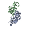
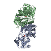
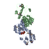
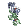
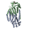
- PDB-3zy6: Crystal structure of POFUT1 in complex with GDP-fucose (crystal-f... -
+ Open data

- Basic information
| Entry | Database: PDB / ID: 3zy6 | ||||||
|---|---|---|---|---|---|---|---|
| Title | Crystal structure of POFUT1 in complex with GDP-fucose (crystal-form-II) | ||||||
 Components Components | PUTATIVE GDP-FUCOSE PROTEIN O-FUCOSYLTRANSFERASE 1 | ||||||
 Keywords Keywords | TRANSFERASE / GLYCOSYLTRANSFERASE / GT-B / CATALYTIC MECHANISM / GT65 | ||||||
| Function / homology |  Function and homology information Function and homology informationpeptide-O-fucosyltransferase / protein O-linked glycosylation via fucose / peptide-O-fucosyltransferase activity / fucose metabolic process / Notch signaling pathway / endoplasmic reticulum Similarity search - Function | ||||||
| Biological species |  | ||||||
| Method |  X-RAY DIFFRACTION / MOLECULAR REPLACEMENT / Resolution: 1.91 Å X-RAY DIFFRACTION / MOLECULAR REPLACEMENT / Resolution: 1.91 Å | ||||||
 Authors Authors | Lira-Navarrete, E. / Valero-Gonzalez, J. / Villanueva, R. / Martinez-Julvez, M. / Tejero, T. / Merino, P. / Panjikar, S. / Hurtado-Guerrero, R. | ||||||
 Citation Citation | Journal: Plos One / Year: 2011 Title: Structural Insights Into the Mechanism of Protein O-Fucosylation. Authors: Lira-Navarrete, E. / Valero-Gonzalez, J. / Villanueva, R. / Martinez-Julvez, M. / Tejero, T. / Merino, P. / Panjikar, S. / Hurtado-Guerrero, R. | ||||||
| History |
|
- Structure visualization
| Structure viewer | Molecule: MolmilJmol/JSmol |
|---|
- Downloads & links
-Download
| PDBx/mmCIF format | 3zy6.cif.gz | 162.9 KB | Display | PDBx/mmCIF format |
|---|---|---|---|---|
| PDB format | pdb3zy6.ent.gz | 127.3 KB | Display | PDB format |
| PDBx/mmJSON format | 3zy6.json.gz | Tree view | PDBx/mmJSON format | |
| Others |  Other downloads Other downloads |
-Validation report
| Arichive directory | https://data.pdbj.org/pub/pdb/validation_reports/zy/3zy6ftp://data.pdbj.org/pub/pdb/validation_reports/zy/3zy6 | HTTPS FTP |
|---|
-Related structure data
| Related structure data |  3zy2SC  3zy3C  3zy4C  3zy5C S: Starting model for refinement C: citing same article ( |
|---|---|
| Similar structure data |
-Links
 PDBj
PDBj- Assembly
| Deposited unit | 
| ||||||||
|---|---|---|---|---|---|---|---|---|---|
| 1 | 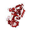
| ||||||||
| Unit cell |
|
-Components
| #1: Protein | Mass: 41008.746 Da / Num. of mol.: 1 / Fragment: RESIDUES 26-383 Source method: isolated from a genetically manipulated source Source: (gene. exp.)  PICHIA PASTORIS (fungus) / Strain (production host): X-33 / References: UniProt: Q18014, peptide-O-fucosyltransferase PICHIA PASTORIS (fungus) / Strain (production host): X-33 / References: UniProt: Q18014, peptide-O-fucosyltransferase |
|---|---|
| #2: Chemical | ChemComp-GFB /   Mass: 589.342 Da / Num. of mol.: 1 / Source method: obtained synthetically / Formula: C16H25N5O15P2 Mass: 589.342 Da / Num. of mol.: 1 / Source method: obtained synthetically / Formula: C16H25N5O15P2 |
| #3: Water | ChemComp-HOH /  Mass: 18.015 Da / Num. of mol.: 243 / Source method: isolated from a natural source / Formula: H2O Mass: 18.015 Da / Num. of mol.: 243 / Source method: isolated from a natural source / Formula: H2O |
| Has protein modification | Y |
-Experimental details
-Experiment
| Experiment | Method: X-RAY DIFFRACTION |
|---|
- Sample preparation
| Crystal | Density Matthews: 2.29 Å3/Da / Density % sol: 46 % / Description: NONE |
|---|
-Data collection
| Diffraction | Mean temperature: 90 K |
|---|---|
| Diffraction source | Source: ROTATING ANODE / Site: ESRF  / Beamline: BM16 / Type: BRUKER / Wavelength: 1.54 / Beamline: BM16 / Type: BRUKER / Wavelength: 1.54 |
| Radiation | Protocol: SINGLE WAVELENGTH / Monochromatic (M) / Laue (L): M / Scattering type: x-ray |
| Radiation wavelength | Wavelength: 1.54 Å / Relative weight: 1 |
| Reflection | Resolution: 1.91→20 Å / Num. obs: 39386 / % possible obs: 98.9 % / Observed criterion σ(I): 2 / Redundancy: 4.37 % / Rmerge(I) obs: 0.1 / Net I/σ(I): 9.27 |
| Reflection shell | Resolution: 1.91→2.01 Å / Redundancy: 3.91 % / Rmerge(I) obs: 0.52 / Mean I/σ(I) obs: 1.97 / % possible all: 96.5 |
- Processing
| Software |
| ||||||||||||||||||||||||||||||||||||||||||||||||||||||||||||||||||||||||||||||||||||||||||||||||||||||||||||||||||||||||||||||||||||||||||||||||||||||||||||||||||||||||||||||||||||||
|---|---|---|---|---|---|---|---|---|---|---|---|---|---|---|---|---|---|---|---|---|---|---|---|---|---|---|---|---|---|---|---|---|---|---|---|---|---|---|---|---|---|---|---|---|---|---|---|---|---|---|---|---|---|---|---|---|---|---|---|---|---|---|---|---|---|---|---|---|---|---|---|---|---|---|---|---|---|---|---|---|---|---|---|---|---|---|---|---|---|---|---|---|---|---|---|---|---|---|---|---|---|---|---|---|---|---|---|---|---|---|---|---|---|---|---|---|---|---|---|---|---|---|---|---|---|---|---|---|---|---|---|---|---|---|---|---|---|---|---|---|---|---|---|---|---|---|---|---|---|---|---|---|---|---|---|---|---|---|---|---|---|---|---|---|---|---|---|---|---|---|---|---|---|---|---|---|---|---|---|---|---|---|---|
| Refinement | Method to determine structure: MOLECULAR REPLACEMENT Starting model: PDB ENTRY 3ZY2 Resolution: 1.91→70.48 Å / Cor.coef. Fo:Fc: 0.945 / Cor.coef. Fo:Fc free: 0.927 / SU B: 8.339 / SU ML: 0.115 / Cross valid method: THROUGHOUT / ESU R: 0.176 / ESU R Free: 0.154 / Stereochemistry target values: MAXIMUM LIKELIHOOD / Details: HYDROGENS HAVE BEEN ADDED IN THE RIDING POSITIONS.
| ||||||||||||||||||||||||||||||||||||||||||||||||||||||||||||||||||||||||||||||||||||||||||||||||||||||||||||||||||||||||||||||||||||||||||||||||||||||||||||||||||||||||||||||||||||||
| Solvent computation | Ion probe radii: 0.8 Å / Shrinkage radii: 0.8 Å / VDW probe radii: 1.4 Å / Solvent model: MASK | ||||||||||||||||||||||||||||||||||||||||||||||||||||||||||||||||||||||||||||||||||||||||||||||||||||||||||||||||||||||||||||||||||||||||||||||||||||||||||||||||||||||||||||||||||||||
| Displacement parameters | Biso mean: 26.879 Å2
| ||||||||||||||||||||||||||||||||||||||||||||||||||||||||||||||||||||||||||||||||||||||||||||||||||||||||||||||||||||||||||||||||||||||||||||||||||||||||||||||||||||||||||||||||||||||
| Refinement step | Cycle: LAST / Resolution: 1.91→70.48 Å
| ||||||||||||||||||||||||||||||||||||||||||||||||||||||||||||||||||||||||||||||||||||||||||||||||||||||||||||||||||||||||||||||||||||||||||||||||||||||||||||||||||||||||||||||||||||||
| Refine LS restraints |
|