 Movie
Movie Controller

[English] 日本語
 Yorodumi
- PDB-1pqu: Crystal Structure of the H277N Mutant of Aspartate Semialdehyde D... -
+ Open data

- Basic information
| Entry | Database: PDB / ID: 1pqu | ||||||
|---|---|---|---|---|---|---|---|
| Title | Crystal Structure of the H277N Mutant of Aspartate Semialdehyde Dehydrogenase from Haemophilus influenzae Bound with NADP, S-methyl cysteine sulfoxide and cacodylate | ||||||
 Components Components | Aspartate-semialdehyde dehydrogenase | ||||||
 Keywords Keywords | OXIDOREDUCTASE / Enzyme / L-aspartate semialdehyde / cacodylate / NADP | ||||||
| Function / homology |  Function and homology information Function and homology informationaspartate-semialdehyde dehydrogenase / aspartate-semialdehyde dehydrogenase activity / 'de novo' L-methionine biosynthetic process / L-threonine biosynthetic process / : / : / : / NAD binding / NADP binding / protein dimerization activity Similarity search - Function | ||||||
| Biological species |  Haemophilus influenzae Rd (bacteria) Haemophilus influenzae Rd (bacteria) | ||||||
| Method |  X-RAY DIFFRACTION / SYNCHROTRON / MOLECULAR REPLACEMENT / Resolution: 1.92 Å X-RAY DIFFRACTION / SYNCHROTRON / MOLECULAR REPLACEMENT / Resolution: 1.92 Å | ||||||
 Authors Authors | Blanco, J. / Moore, R.A. / Viola, R.E. | ||||||
 Citation Citation | Journal: Acta Crystallogr.,Sect.D / Year: 2004 Title: Critical catalytic functional groups in the mechanism of aspartate-beta-semialdehyde dehydrogenase. Authors: Blanco, J. / Moore, R.A. / Faehnle, C.R. / Viola, R.E. #1: Journal: Acta Crystallogr.,Sect.D / Year: 2004Title: The role of substrate-binding groups in the mechanism of aspartate-beta-semialdehyde dehydrogenase. Authors: Blanco, J. / Moore, R.A. / Faehnle, C.R. / Coe, D.M. / Viola, R.E. | ||||||
| History |
|
- Structure visualization
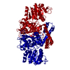
| Structure viewer | Molecule: MolmilJmol/JSmol |
|---|
- Downloads & links
-Download
| PDBx/mmCIF format | 1pqu.cif.gz | 307.5 KB | Display | PDBx/mmCIF format |
|---|---|---|---|---|
| PDB format | pdb1pqu.ent.gz | 248.7 KB | Display | PDB format |
| PDBx/mmJSON format | 1pqu.json.gz | Tree view | PDBx/mmJSON format | |
| Others |  Other downloads Other downloads |
-Validation report
| Arichive directory | https://data.pdbj.org/pub/pdb/validation_reports/pq/1pquftp://data.pdbj.org/pub/pdb/validation_reports/pq/1pqu | HTTPS FTP |
|---|
-Related structure data
| Related structure data |  1pqpC  1nwcS S: Starting model for refinement C: citing same article ( |
|---|---|
| Similar structure data |


-Links
 PDBj
PDBj


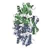
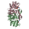
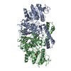
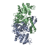
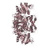
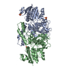
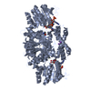
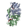
- Assembly
| Deposited unit | 
| ||||||||
|---|---|---|---|---|---|---|---|---|---|
| 1 | 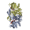
| ||||||||
| 2 | 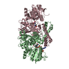
| ||||||||
| Unit cell |
|
-Components
| #1: Protein | Mass: 40559.691 Da / Num. of mol.: 4 / Mutation: H277N Source method: isolated from a genetically manipulated source Source: (gene. exp.) Haemophilus influenzae Rd (bacteria) / Species: Haemophilus influenzae / Strain: KW20 / Gene: asd / Plasmid: PET43 / Species (production host): Escherichia coli / Production host: References: UniProt: P44801, aspartate-semialdehyde dehydrogenase #2: Chemical | ChemComp-CAC /   Mass: 136.989 Da / Num. of mol.: 4 / Source method: obtained synthetically / Formula: C2H6AsO2 Mass: 136.989 Da / Num. of mol.: 4 / Source method: obtained synthetically / Formula: C2H6AsO2#3: Chemical |   Mass: 743.405 Da / Num. of mol.: 3 / Source method: obtained synthetically / Formula: C21H28N7O17P3 Mass: 743.405 Da / Num. of mol.: 3 / Source method: obtained synthetically / Formula: C21H28N7O17P3#4: Chemical | ChemComp-CYS /   Type: L-peptide linking / Mass: 121.158 Da / Num. of mol.: 4 / Source method: obtained synthetically / Formula: C3H7NO2S Type: L-peptide linking / Mass: 121.158 Da / Num. of mol.: 4 / Source method: obtained synthetically / Formula: C3H7NO2S#5: Water | ChemComp-HOH / |  Mass: 18.015 Da / Num. of mol.: 799 / Source method: isolated from a natural source / Formula: H2O Mass: 18.015 Da / Num. of mol.: 799 / Source method: isolated from a natural source / Formula: H2OHas protein modification | Y | |
|---|
-Experimental details
-Experiment
| Experiment | Method: X-RAY DIFFRACTION / Number of used crystals: 1 |
|---|
- Sample preparation
| Crystal | Density Matthews: 2.3 Å3/Da / Density % sol: 46.63 % |
|---|---|
| Crystal grow | Temperature: 298 K / Method: vapor diffusion, hanging drop / pH: 6.5 Details: 24% PEG 3350, 0.2M Ammonium acetate, 100 mM cacodylate, pH 6.5, 2 mM NADP, 2 mM S-methyl cysteine sulfoxide, VAPOR DIFFUSION, HANGING DROP, temperature 298K |
-Data collection
| Diffraction | Mean temperature: 90 K |
|---|---|
| Diffraction source | Source: SYNCHROTRON / Site: APS  / Beamline: 19-BM / Wavelength: 1 Å / Beamline: 19-BM / Wavelength: 1 Å |
| Detector | Type: SBC-3 / Detector: CCD / Date: Jul 18, 2002 |
| Radiation | Monochromator: Si220 / Protocol: SINGLE WAVELENGTH / Monochromatic (M) / Laue (L): M / Scattering type: x-ray |
| Radiation wavelength | Wavelength: 1 Å / Relative weight: 1 |
| Reflection | Resolution: 1.92→50 Å / Num. obs: 99660 / % possible obs: 89 % / Observed criterion σ(F): 2 / Observed criterion σ(I): 2 / Redundancy: 7.4 % / Biso Wilson estimate: 9.9 Å2 / Rsym value: 0.08 / Net I/σ(I): 10.7 |
| Reflection shell | Resolution: 1.92→1.99 Å / Mean I/σ(I) obs: 2.5 / Num. unique all: 10047 / Rsym value: 0.225 / % possible all: 90.7 |
- Processing
| Software |
| |||||||||||||||||||||||||
|---|---|---|---|---|---|---|---|---|---|---|---|---|---|---|---|---|---|---|---|---|---|---|---|---|---|---|
| Refinement | Method to determine structure: MOLECULAR REPLACEMENT Starting model: 1NWC Resolution: 1.92→44.65 Å / Rfactor Rfree error: 0.002 / Isotropic thermal model: RESTRAINED / Cross valid method: THROUGHOUT / σ(F): 0 / σ(I): 2 / Stereochemistry target values: Engh & Huber
| |||||||||||||||||||||||||
| Solvent computation | Solvent model: FLAT MODEL / Bsol: 59.5703 Å2 / ksol: 0.384788 e/Å3 | |||||||||||||||||||||||||
| Displacement parameters | Biso mean: 18.4 Å2
| |||||||||||||||||||||||||
| Refine analyze |
| |||||||||||||||||||||||||
| Refinement step | Cycle: LAST / Resolution: 1.92→44.65 Å
| |||||||||||||||||||||||||
| Refine LS restraints |
| |||||||||||||||||||||||||
| LS refinement shell | Resolution: 1.92→2.04 Å / Rfactor Rfree error: 0.007 / Total num. of bins used: 6
| |||||||||||||||||||||||||
| Xplor file |
|
