 Movie
Movie Controller

[English] 日本語
 Yorodumi
- PDB-1mb4: Crystal structure of aspartate semialdehyde dehydrogenase from vi... -
+ Open data

- Basic information
| Entry | Database: PDB / ID: 1mb4 | ||||||
|---|---|---|---|---|---|---|---|
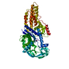
| Title | Crystal structure of aspartate semialdehyde dehydrogenase from vibrio cholerae with NADP and S-methyl-l-cysteine sulfoxide | ||||||
 Components Components | Aspartate-Semialdehyde Dehydrogenase | ||||||
 Keywords Keywords | OXIDOREDUCTASE / enzyme / aspartate-semialdehyde dehydrogenase / complex / vibrio cholerae | ||||||
| Function / homology |  Function and homology information Function and homology informationaspartate-semialdehyde dehydrogenase / aspartate-semialdehyde dehydrogenase activity / 'de novo' L-methionine biosynthetic process / L-threonine biosynthetic process / : / : / : / NAD binding / NADP binding / protein dimerization activity Similarity search - Function | ||||||
| Biological species |   Vibrio cholerae (bacteria) Vibrio cholerae (bacteria) | ||||||
| Method |  X-RAY DIFFRACTION / SYNCHROTRON / MOLECULAR REPLACEMENT / Resolution: 1.84 Å X-RAY DIFFRACTION / SYNCHROTRON / MOLECULAR REPLACEMENT / Resolution: 1.84 Å | ||||||
 Authors Authors | Blanco, J. / Moore, R.A. / Kabaleeswaran, V. / Viola, R.E. | ||||||
 Citation Citation | Journal: Protein Sci. / Year: 2003 Title: A structural Basis for the Mechanism of Aspartate-beta-semialdehyde Dehydrogenase from Vibrio Cholerae Authors: Blanco, J. / Moore, R.A. / Kabaleeswaran, V. / Viola, R.E. | ||||||
| History |
|
- Structure visualization
| Structure viewer | Molecule: MolmilJmol/JSmol |
|---|
- Downloads & links
-Download
| PDBx/mmCIF format | 1mb4.cif.gz | 162 KB | Display | PDBx/mmCIF format |
|---|---|---|---|---|
| PDB format | pdb1mb4.ent.gz | 128.5 KB | Display | PDB format |
| PDBx/mmJSON format | 1mb4.json.gz | Tree view | PDBx/mmJSON format | |
| Others |  Other downloads Other downloads |
-Validation report
| Arichive directory | https://data.pdbj.org/pub/pdb/validation_reports/mb/1mb4ftp://data.pdbj.org/pub/pdb/validation_reports/mb/1mb4 | HTTPS FTP |
|---|
-Related structure data
| Related structure data |  1mc4C  1gl3S S: Starting model for refinement C: citing same article ( |
|---|---|
| Similar structure data |


-Links
 PDBj
PDBj


- Assembly
| Deposited unit | 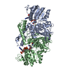
| ||||||||
|---|---|---|---|---|---|---|---|---|---|
| 1 |
| ||||||||
| Unit cell |
| ||||||||
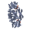
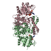
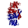
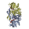
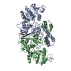
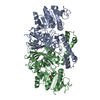
| Details | The biological assembly is a homodimer |
-Components
| #1: Protein | Mass: 40541.664 Da / Num. of mol.: 2 Source method: isolated from a genetically manipulated source Source: (gene. exp.) Vibrio cholerae (bacteria) / Gene: asd / Plasmid: PET41 / Species (production host): Escherichia coli / Production host: References: UniProt: Q9KQG2, aspartate-semialdehyde dehydrogenase #2: Chemical |   Type: L-peptide linking / Mass: 121.158 Da / Num. of mol.: 2 / Source method: obtained synthetically / Formula: C3H7NO2S Type: L-peptide linking / Mass: 121.158 Da / Num. of mol.: 2 / Source method: obtained synthetically / Formula: C3H7NO2S#3: Chemical |   Mass: 745.421 Da / Num. of mol.: 2 / Source method: obtained synthetically / Formula: C21H30N7O17P3 Mass: 745.421 Da / Num. of mol.: 2 / Source method: obtained synthetically / Formula: C21H30N7O17P3#4: Water | ChemComp-HOH / |  Mass: 18.015 Da / Num. of mol.: 457 / Source method: isolated from a natural source / Formula: H2O Mass: 18.015 Da / Num. of mol.: 457 / Source method: isolated from a natural source / Formula: H2OHas protein modification | Y | |
|---|
-Experimental details
-Experiment
| Experiment | Method: X-RAY DIFFRACTION / Number of used crystals: 1 |
|---|
- Sample preparation
| Crystal | Density Matthews: 2.74 Å3/Da / Density % sol: 55.14 % | ||||||||||||||||||||||||||||||||||||||||||
|---|---|---|---|---|---|---|---|---|---|---|---|---|---|---|---|---|---|---|---|---|---|---|---|---|---|---|---|---|---|---|---|---|---|---|---|---|---|---|---|---|---|---|---|
| Crystal grow | Temperature: 293 K / Method: vapor diffusion, hanging drop / pH: 5.6 Details: 20% PEG 3350, 0.2M Sodium Acetate, 0.1M Sodium citrate pH 5.6, VAPOR DIFFUSION, HANGING DROP | ||||||||||||||||||||||||||||||||||||||||||
| Crystal grow | *PLUS | ||||||||||||||||||||||||||||||||||||||||||
| Components of the solutions | *PLUS
|
-Data collection
| Diffraction | Mean temperature: 90 K |
|---|---|
| Diffraction source | Source: SYNCHROTRON / Site: APS  / Beamline: 14-BM-C / Wavelength: 1 Å / Beamline: 14-BM-C / Wavelength: 1 Å |
| Detector | Type: ADSC QUANTUM 4 / Detector: CCD / Date: Sep 1, 2001 |
| Radiation | Monochromator: Diamond (111) double-crystal / Protocol: SINGLE WAVELENGTH / Monochromatic (M) / Laue (L): M / Scattering type: x-ray |
| Radiation wavelength | Wavelength: 1 Å / Relative weight: 1 |
| Reflection | Resolution: 1.84→62.5 Å / Num. obs: 76343 / % possible obs: 99.5 % / Observed criterion σ(F): 1 / Observed criterion σ(I): 1 / Redundancy: 12.2 % / Biso Wilson estimate: 16.8 Å2 / Rmerge(I) obs: 0.084 / Rsym value: 0.084 / Net I/σ(I): 19.4 |
| Reflection shell | Resolution: 1.84→1.91 Å / Redundancy: 10.45 % / Rmerge(I) obs: 0.34 / Mean I/σ(I) obs: 3.59 / Num. unique all: 14265 / Rsym value: 0.34 / % possible all: 95.4 |
| Reflection | *PLUS Lowest resolution: 50 Å / Num. obs: 149813 / Num. measured all: 1811452 |
| Reflection shell | *PLUS % possible obs: 95.4 % |
- Processing
| Software |
| ||||||||||||||||||||||||||||||||||||||||||||||||||||||||||||||||||||||||||||||||
|---|---|---|---|---|---|---|---|---|---|---|---|---|---|---|---|---|---|---|---|---|---|---|---|---|---|---|---|---|---|---|---|---|---|---|---|---|---|---|---|---|---|---|---|---|---|---|---|---|---|---|---|---|---|---|---|---|---|---|---|---|---|---|---|---|---|---|---|---|---|---|---|---|---|---|---|---|---|---|---|---|---|
| Refinement | Method to determine structure: MOLECULAR REPLACEMENT Starting model: PDB ENTRY 1GL3 Resolution: 1.84→62.5 Å / Rfactor Rfree error: 0.003 / Data cutoff high absF: 468178.78 / Data cutoff high rms absF: 468178.78 / Isotropic thermal model: RESTRAINED / Cross valid method: THROUGHOUT / σ(F): 0 / σ(I): 0 / Stereochemistry target values: CNS
| ||||||||||||||||||||||||||||||||||||||||||||||||||||||||||||||||||||||||||||||||
| Solvent computation | Solvent model: FLAT MODEL / Bsol: 67.1137 Å2 / ksol: 0.376184 e/Å3 | ||||||||||||||||||||||||||||||||||||||||||||||||||||||||||||||||||||||||||||||||
| Displacement parameters | Biso mean: 23.3 Å2
| ||||||||||||||||||||||||||||||||||||||||||||||||||||||||||||||||||||||||||||||||
| Refine analyze |
| ||||||||||||||||||||||||||||||||||||||||||||||||||||||||||||||||||||||||||||||||
| Refinement step | Cycle: LAST / Resolution: 1.84→62.5 Å
| ||||||||||||||||||||||||||||||||||||||||||||||||||||||||||||||||||||||||||||||||
| Refine LS restraints |
| ||||||||||||||||||||||||||||||||||||||||||||||||||||||||||||||||||||||||||||||||
| LS refinement shell | Refine-ID: X-RAY DIFFRACTION / Total num. of bins used: 6
| ||||||||||||||||||||||||||||||||||||||||||||||||||||||||||||||||||||||||||||||||
| Xplor file |
| ||||||||||||||||||||||||||||||||||||||||||||||||||||||||||||||||||||||||||||||||
| Refinement | *PLUS Lowest resolution: 50 Å / % reflection Rfree: 5 % | ||||||||||||||||||||||||||||||||||||||||||||||||||||||||||||||||||||||||||||||||
| Solvent computation | *PLUS | ||||||||||||||||||||||||||||||||||||||||||||||||||||||||||||||||||||||||||||||||
| Displacement parameters | *PLUS | ||||||||||||||||||||||||||||||||||||||||||||||||||||||||||||||||||||||||||||||||
| Refine LS restraints | *PLUS
|
