 Movie
Movie Controller

[English] 日本語
 Yorodumi
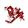
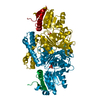
- PDB-1gl3: ASPARTATE BETA-SEMIALDEHYDE DEHYDROGENASE IN COMPLEX WITH NADP AN... -
+ Open data

- Basic information
| Entry | Database: PDB / ID: 1gl3 | ||||||
|---|---|---|---|---|---|---|---|
| Title | ASPARTATE BETA-SEMIALDEHYDE DEHYDROGENASE IN COMPLEX WITH NADP AND SUBSTRATE ANALOGUE S-METHYL CYSTEINE SULFOXIDE | ||||||
 Components Components | ASPARTATE-SEMIALDEHYDE DEHYDROGENASE | ||||||
 Keywords Keywords | OXIDOREDUCTASE / DEHYDROGENASE / ESCHERICHIA COLI / ENZYME / NADP / DIAMINOPIMELATE BIOSYNTHESI LYSINE BIOSYNTHESIS | ||||||
| Function / homology |  Function and homology information Function and homology informationL-homoserine biosynthetic process / aspartate-semialdehyde dehydrogenase / aspartate-semialdehyde dehydrogenase activity / 'de novo' L-methionine biosynthetic process / L-threonine biosynthetic process / : / : / : / NAD binding / NADP binding ...L-homoserine biosynthetic process / aspartate-semialdehyde dehydrogenase / aspartate-semialdehyde dehydrogenase activity / 'de novo' L-methionine biosynthetic process / L-threonine biosynthetic process / : / : / : / NAD binding / NADP binding / protein dimerization activity / DNA damage response / cytosol Similarity search - Function | ||||||
| Biological species |  | ||||||
| Method |  X-RAY DIFFRACTION / MOLECULAR REPLACEMENT / Resolution: 2.6 Å X-RAY DIFFRACTION / MOLECULAR REPLACEMENT / Resolution: 2.6 Å | ||||||
 Authors Authors | Hadfield, A.T. / Kryger, G. / Ouyang, J. / Ringe, D. / Petsko, G.A. / Viola, R.E. | ||||||
 Citation Citation | Journal: Biochemistry / Year: 2001 Title: Active Site Analysis of the Potential Antimicrobial Target Aspartate Semialdehyde Dehydrogenase. Authors: Hadfield, A.T. / Shammas, C. / Kryger, G. / Ringe, D. / Petsko, G.A. / Ouyang, J. / Viola, R.E. #1: Journal: J.Mol.Biol. / Year: 1999Title: Structure of Aspartate-Beta-Semialdehyde Dehydrogenase from Escherichia Coli, a Key Enzyme in the Aspartate Family of Amino Acid Biosynthesis Authors: Hadfield, A.T. / Kryger, G. / Ouyang, J. / Petsko, G.A. / Viola, R.E. | ||||||
| History |
|
- Structure visualization
| Structure viewer | Molecule: MolmilJmol/JSmol |
|---|
- Downloads & links
-Download
| PDBx/mmCIF format | 1gl3.cif.gz | 153.7 KB | Display | PDBx/mmCIF format |
|---|---|---|---|---|
| PDB format | pdb1gl3.ent.gz | 122 KB | Display | PDB format |
| PDBx/mmJSON format | 1gl3.json.gz | Tree view | PDBx/mmJSON format | |
| Others |  Other downloads Other downloads |
-Validation report
| Arichive directory | https://data.pdbj.org/pub/pdb/validation_reports/gl/1gl3ftp://data.pdbj.org/pub/pdb/validation_reports/gl/1gl3 | HTTPS FTP |
|---|
-Related structure data
| Related structure data |  1brmS S: Starting model for refinement |
|---|---|
| Similar structure data |


-Links
 PDBj
PDBj


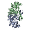
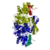
- Assembly
| Deposited unit | 
| ||||||||
|---|---|---|---|---|---|---|---|---|---|
| 1 |
| ||||||||
| Unit cell |
| ||||||||
| Noncrystallographic symmetry (NCS) | NCS oper: (Code: given Matrix: (-0.9746, 0.224, -0.0013), Vector: |
-Components
| #1: Protein | Mass: 40056.738 Da / Num. of mol.: 2 Source method: isolated from a genetically manipulated source Source: (gene. exp.) References: UniProt: P00353, UniProt: P0A9Q9*PLUS, aspartate-semialdehyde dehydrogenase #2: Chemical | ChemComp-CYS / |   Type: L-peptide linking / Mass: 121.158 Da / Num. of mol.: 1 / Source method: obtained synthetically / Formula: C3H7NO2S Type: L-peptide linking / Mass: 121.158 Da / Num. of mol.: 1 / Source method: obtained synthetically / Formula: C3H7NO2S#3: Chemical |   Mass: 745.421 Da / Num. of mol.: 2 / Source method: obtained synthetically / Formula: C21H30N7O17P3 Mass: 745.421 Da / Num. of mol.: 2 / Source method: obtained synthetically / Formula: C21H30N7O17P3#4: Water | ChemComp-HOH / |  Mass: 18.015 Da / Num. of mol.: 171 / Source method: isolated from a natural source / Formula: H2O Mass: 18.015 Da / Num. of mol.: 171 / Source method: isolated from a natural source / Formula: H2OHas protein modification | Y | |
|---|
-Experimental details
-Experiment
| Experiment | Method: X-RAY DIFFRACTION / Number of used crystals: 1 |
|---|
- Sample preparation
| Crystal | Density Matthews: 2.75 Å3/Da / Density % sol: 55 % | ||||||||||||||||||||||||||||||||||||||||||||||||||||||||||||||||||||||||||||||||||||
|---|---|---|---|---|---|---|---|---|---|---|---|---|---|---|---|---|---|---|---|---|---|---|---|---|---|---|---|---|---|---|---|---|---|---|---|---|---|---|---|---|---|---|---|---|---|---|---|---|---|---|---|---|---|---|---|---|---|---|---|---|---|---|---|---|---|---|---|---|---|---|---|---|---|---|---|---|---|---|---|---|---|---|---|---|---|
| Crystal grow | pH: 4.6 Details: RESERVOIR: 19% PEG 4K, 0.35M MGCL2, 0.2 M NH4AC 0.1M TRIS, pH 4.60 | ||||||||||||||||||||||||||||||||||||||||||||||||||||||||||||||||||||||||||||||||||||
| Crystal grow | *PLUS Temperature: 4 ℃ / Method: vapor diffusion, hanging drop / pH: 7 | ||||||||||||||||||||||||||||||||||||||||||||||||||||||||||||||||||||||||||||||||||||
| Components of the solutions | *PLUS
|
-Data collection
| Diffraction | Mean temperature: 100 K |
|---|---|
| Diffraction source | Source: ROTATING ANODE / Type: RIGAKU RU200 / Wavelength: 1.54 |
| Detector | Type: MAR scanner 300 mm plate / Detector: IMAGE PLATE / Date: Oct 15, 1996 |
| Radiation | Monochromator: SI / Protocol: SINGLE WAVELENGTH / Monochromatic (M) / Laue (L): M / Scattering type: x-ray |
| Radiation wavelength | Wavelength: 1.54 Å / Relative weight: 1 |
| Reflection | Resolution: 2.6→28 Å / Num. obs: 41666 / % possible obs: 96.3 % / Observed criterion σ(I): 0 / Redundancy: 1.9 % / Rmerge(I) obs: 0.103 |
| Reflection shell | Resolution: 2.6→2.74 Å / Redundancy: 1.4 % / Rmerge(I) obs: 0.307 / Mean I/σ(I) obs: 1.5 / % possible all: 61.6 |
| Reflection | *PLUS Num. obs: 22099 / Num. measured all: 41666 |
| Reflection shell | *PLUS % possible obs: 61.6 % |
- Processing
| Software |
| ||||||||||||||||||||||||||||||||||||||||||||||||||||||||||||
|---|---|---|---|---|---|---|---|---|---|---|---|---|---|---|---|---|---|---|---|---|---|---|---|---|---|---|---|---|---|---|---|---|---|---|---|---|---|---|---|---|---|---|---|---|---|---|---|---|---|---|---|---|---|---|---|---|---|---|---|---|---|
| Refinement | Method to determine structure: MOLECULAR REPLACEMENT Starting model: PDB ENTRY 1BRM Resolution: 2.6→28 Å / Data cutoff high absF: 100000 / Data cutoff low absF: 0.001 / Isotropic thermal model: RESTRAINED / Cross valid method: THROUGHOUT / σ(F): 0
| ||||||||||||||||||||||||||||||||||||||||||||||||||||||||||||
| Displacement parameters | Biso mean: 29.1 Å2 | ||||||||||||||||||||||||||||||||||||||||||||||||||||||||||||
| Refinement step | Cycle: LAST / Resolution: 2.6→28 Å
| ||||||||||||||||||||||||||||||||||||||||||||||||||||||||||||
| Refine LS restraints |
| ||||||||||||||||||||||||||||||||||||||||||||||||||||||||||||
| LS refinement shell | Resolution: 2.6→2.74 Å / Total num. of bins used: 8
| ||||||||||||||||||||||||||||||||||||||||||||||||||||||||||||
| Xplor file |
| ||||||||||||||||||||||||||||||||||||||||||||||||||||||||||||
| Software | *PLUS Name: X-PLOR / Version: 3.85 / Classification: refinement | ||||||||||||||||||||||||||||||||||||||||||||||||||||||||||||
| Refinement | *PLUS Rfactor obs: 0.2 / Rfactor Rwork: 0.2 | ||||||||||||||||||||||||||||||||||||||||||||||||||||||||||||
| Solvent computation | *PLUS | ||||||||||||||||||||||||||||||||||||||||||||||||||||||||||||
| Displacement parameters | *PLUS Biso mean: 28.3 Å2 |
