 Movie
Movie Controller

[English] 日本語
 Yorodumi
- PDB-3vos: Crystal structure of Aspartate semialdehyde dehydrogenase Complex... -
+ Open data

- Basic information
| Entry | Database: PDB / ID: 3vos | |||||||||
|---|---|---|---|---|---|---|---|---|---|---|
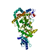
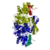
| Title | Crystal structure of Aspartate semialdehyde dehydrogenase Complexed With glycerol and sulfate From Mycobacterium tuberculosis H37Rv | |||||||||
 Components Components | Aspartate-semialdehyde dehydrogenase | |||||||||
 Keywords Keywords | OXIDOREDUCTASE / DEHYDROGENASE / AMINO-ACID BIOSYNTHESIS / LYSINE BIOSYNTHESIS | |||||||||
| Function / homology |  Function and homology information Function and homology informationaspartate-semialdehyde dehydrogenase / aspartate-semialdehyde dehydrogenase activity / 'de novo' L-methionine biosynthetic process / cell wall / L-threonine biosynthetic process / : / : / : / peptidoglycan-based cell wall / NAD binding ...aspartate-semialdehyde dehydrogenase / aspartate-semialdehyde dehydrogenase activity / 'de novo' L-methionine biosynthetic process / cell wall / L-threonine biosynthetic process / : / : / : / peptidoglycan-based cell wall / NAD binding / NADP binding / protein dimerization activity / plasma membrane Similarity search - Function | |||||||||
| Biological species |   Mycobacterium tuberculosis (bacteria) Mycobacterium tuberculosis (bacteria) | |||||||||
| Method |  X-RAY DIFFRACTION / SYNCHROTRON / MOLECULAR REPLACEMENT / Resolution: 2.18 Å X-RAY DIFFRACTION / SYNCHROTRON / MOLECULAR REPLACEMENT / Resolution: 2.18 Å | |||||||||
 Authors Authors | Vyas, R. / Tewari, R. / Weiss, M.S. / Karthikeyan, S. | |||||||||
 Citation Citation | Journal: Acta Crystallogr.,Sect.D / Year: 2012 Title: Structures of ternary complexes of aspartate-semialdehyde dehydrogenase (Rv3708c) from Mycobacterium tuberculosis H37Rv Authors: Vyas, R. / Tewari, R. / Weiss, M.S. / Karthikeyan, S. | |||||||||
| History |
|
- Structure visualization
| Structure viewer | Molecule: MolmilJmol/JSmol |
|---|
- Downloads & links
-Download
| PDBx/mmCIF format | 3vos.cif.gz | 85.4 KB | Display | PDBx/mmCIF format |
|---|---|---|---|---|
| PDB format | pdb3vos.ent.gz | 65 KB | Display | PDB format |
| PDBx/mmJSON format | 3vos.json.gz | Tree view | PDBx/mmJSON format | |
| Others |  Other downloads Other downloads |
-Validation report
| Arichive directory | https://data.pdbj.org/pub/pdb/validation_reports/vo/3vosftp://data.pdbj.org/pub/pdb/validation_reports/vo/3vos | HTTPS FTP |
|---|
-Related structure data
| Related structure data |  3tz6C  2gyyS C: citing same article ( S: Starting model for refinement |
|---|---|
| Similar structure data |


-Links
 PDBj
PDBj- Assembly
| Deposited unit | 
| ||||||||
|---|---|---|---|---|---|---|---|---|---|
| 1 | 
| ||||||||
| Unit cell |
| ||||||||
| Components on special symmetry positions |
|
-Components
| #1: Protein | Mass: 38114.008 Da / Num. of mol.: 1 Source method: isolated from a genetically manipulated source Source: (gene. exp.) Mycobacterium tuberculosis (bacteria) / Strain: h37rv / Gene: asd, MT3811, MTV025.056c, Rv3708c / Plasmid: pQE-30 / Production host: References: UniProt: P0A542, UniProt: P9WNX5*PLUS, aspartate-semialdehyde dehydrogenase | ||||
|---|---|---|---|---|---|
| #2: Chemical |   Mass: 92.094 Da / Num. of mol.: 3 / Source method: obtained synthetically / Formula: C3H8O3 Mass: 92.094 Da / Num. of mol.: 3 / Source method: obtained synthetically / Formula: C3H8O3#3: Chemical | ChemComp-SO4 /   Mass: 96.063 Da / Num. of mol.: 6 / Source method: obtained synthetically / Formula: SO4 Mass: 96.063 Da / Num. of mol.: 6 / Source method: obtained synthetically / Formula: SO4#4: Water | ChemComp-HOH / |  Mass: 18.015 Da / Num. of mol.: 342 / Source method: isolated from a natural source / Formula: H2O Mass: 18.015 Da / Num. of mol.: 342 / Source method: isolated from a natural source / Formula: H2O |
-Experimental details
-Experiment
| Experiment | Method: X-RAY DIFFRACTION / Number of used crystals: 1 |
|---|
- Sample preparation
| Crystal | Density Matthews: 5.24 Å3/Da / Density % sol: 76.5 % |
|---|---|
| Crystal grow | Temperature: 293 K / Method: vapor diffusion, sitting drop / pH: 5 Details: 100MM CITRIC ACID, 1.6M AMSO4, pH 5.0, VAPOR DIFFUSION, SITTING DROP, temperature 293K |
-Data collection
| Diffraction | Mean temperature: 100 K |
|---|---|
| Diffraction source | Source: SYNCHROTRON / Site: ESRF  / Beamline: BM14 / Wavelength: 1.067 Å / Beamline: BM14 / Wavelength: 1.067 Å |
| Detector | Type: MARMOSAIC 225 mm CCD / Detector: CCD / Date: Sep 10, 2007 / Details: Mirrors |
| Radiation | Monochromator: Mirrors / Protocol: SINGLE WAVELENGTH / Monochromatic (M) / Laue (L): M / Scattering type: x-ray |
| Radiation wavelength | Wavelength: 1.067 Å / Relative weight: 1 |
| Reflection | Resolution: 2.18→99 Å / Num. all: 43078 / Num. obs: 43078 / % possible obs: 99.9 % / Observed criterion σ(F): 2.18 / Observed criterion σ(I): -3 / Redundancy: 8.2 % / Biso Wilson estimate: 32.1 Å2 / Rmerge(I) obs: 0.154 / Rsym value: 0.154 / Net I/σ(I): 14.1 |
| Reflection shell | Resolution: 2.18→2.22 Å / % possible obs: 100 % / Redundancy: 8.2 % / Rmerge(I) obs: 0.953 / Mean I/σ(I) obs: 2.2 / Rsym value: 0.953 |
- Processing
| Software |
| |||||||||||||||||||||||||||||||||||||||||||||
|---|---|---|---|---|---|---|---|---|---|---|---|---|---|---|---|---|---|---|---|---|---|---|---|---|---|---|---|---|---|---|---|---|---|---|---|---|---|---|---|---|---|---|---|---|---|---|
| Refinement | Method to determine structure: MOLECULAR REPLACEMENT Starting model: 2GYY Resolution: 2.18→32.42 Å / Cor.coef. Fo:Fc: 0.955 / Cor.coef. Fo:Fc free: 0.952 / SU B: 3.014 / SU ML: 0.076 / Cross valid method: THROUGHOUT / ESU R: 0.125 / ESU R Free: 0.114 / Stereochemistry target values: MAXIMUM LIKELIHOOD / Details: HYDROGENS HAVE BEEN USED IF PRESENT IN THE INPUT
| |||||||||||||||||||||||||||||||||||||||||||||
| Solvent computation | Ion probe radii: 0.8 Å / Shrinkage radii: 0.8 Å / VDW probe radii: 1.2 Å / Solvent model: MASK | |||||||||||||||||||||||||||||||||||||||||||||
| Displacement parameters | Biso mean: 30.848 Å2 | |||||||||||||||||||||||||||||||||||||||||||||
| Refinement step | Cycle: LAST / Resolution: 2.18→32.42 Å
| |||||||||||||||||||||||||||||||||||||||||||||
| Refine LS restraints |
| |||||||||||||||||||||||||||||||||||||||||||||
| LS refinement shell | Resolution: 2.18→2.237 Å / Total num. of bins used: 20
|
