 Movie
Movie Controller

[English] 日本語
 Yorodumi
- PDB-1nx6: Crystal Structure of Aspartate Semialdehyde Dehydrogenase from Ha... -
+ Open data

- Basic information
| Entry | Database: PDB / ID: 1nx6 | ||||||
|---|---|---|---|---|---|---|---|
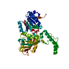
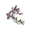
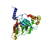
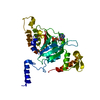
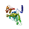
| Title | Crystal Structure of Aspartate Semialdehyde Dehydrogenase from Haemophilus influenzae as a Tetrahedral Hemithiocetal Reaction intermediate with Phosphate at 2.15 A | ||||||
 Components Components | Aspartate-Semialdehyde Dehydrogenase | ||||||
 Keywords Keywords | OXIDOREDUCTASE / aspartate-semialdehyde dehydrogenase / aspartate semialdehyde / Haemophilus influenzae / tetrahedal intermediate / hemithiocetal / phosphate | ||||||
| Function / homology |  Function and homology information Function and homology informationaspartate-semialdehyde dehydrogenase / aspartate-semialdehyde dehydrogenase activity / 'de novo' L-methionine biosynthetic process / L-threonine biosynthetic process / : / : / : / NAD binding / NADP binding / protein dimerization activity Similarity search - Function | ||||||
| Biological species |  Haemophilus influenzae (bacteria) Haemophilus influenzae (bacteria) | ||||||
| Method |  X-RAY DIFFRACTION / SYNCHROTRON / MOLECULAR REPLACEMENT / Resolution: 2.15 Å X-RAY DIFFRACTION / SYNCHROTRON / MOLECULAR REPLACEMENT / Resolution: 2.15 Å | ||||||
 Authors Authors | Blanco, J. / Moore, R.A. / Viola, R.E. | ||||||
 Citation Citation | Journal: Proc.Natl.Acad.Sci.USA / Year: 2003 Title: Capture of an Intermediate in the Catalytic Cycle of L-Aspartate-beta-Semialdehyde Dehydrogenase Authors: Blanco, J. / Moore, R.A. / Viola, R.E. | ||||||
| History |
|
- Structure visualization
| Structure viewer | Molecule: MolmilJmol/JSmol |
|---|
- Downloads & links
-Download
| PDBx/mmCIF format | 1nx6.cif.gz | 85.7 KB | Display | PDBx/mmCIF format |
|---|---|---|---|---|
| PDB format | pdb1nx6.ent.gz | 63.9 KB | Display | PDB format |
| PDBx/mmJSON format | 1nx6.json.gz | Tree view | PDBx/mmJSON format | |
| Others |  Other downloads Other downloads |
-Validation report
| Arichive directory | https://data.pdbj.org/pub/pdb/validation_reports/nx/1nx6ftp://data.pdbj.org/pub/pdb/validation_reports/nx/1nx6 | HTTPS FTP |
|---|
-Related structure data
| Related structure data |  1nwcC  1nwhSC S: Starting model for refinement C: citing same article ( |
|---|---|
| Similar structure data |


-Links
 PDBj
PDBj- Assembly
| Deposited unit | 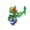
| ||||||||
|---|---|---|---|---|---|---|---|---|---|
| 1 |
| ||||||||
| 2 | 
| ||||||||
| Unit cell |
| ||||||||
| Details | The biological assembly is a monomer |
-Components
| #1: Protein | Mass: 40700.836 Da / Num. of mol.: 1 Source method: isolated from a genetically manipulated source Source: (gene. exp.) Haemophilus influenzae (bacteria) / Gene: asd / Plasmid: PET41a / Species (production host): Escherichia coli / Production host: References: UniProt: P44801, aspartate-semialdehyde dehydrogenase | ||
|---|---|---|---|
| #2: Chemical |   Mass: 94.971 Da / Num. of mol.: 2 / Source method: obtained synthetically / Formula: PO4 Mass: 94.971 Da / Num. of mol.: 2 / Source method: obtained synthetically / Formula: PO4#3: Water | ChemComp-HOH / |  Mass: 18.015 Da / Num. of mol.: 144 / Source method: isolated from a natural source / Formula: H2O Mass: 18.015 Da / Num. of mol.: 144 / Source method: isolated from a natural source / Formula: H2O |
-Experimental details
-Experiment
| Experiment | Method: X-RAY DIFFRACTION / Number of used crystals: 1 |
|---|
- Sample preparation
| Crystal | Density Matthews: 2.21 Å3/Da / Density % sol: 43.78 % | ||||||||||||||||||||||||||||||||||||||||||||||||
|---|---|---|---|---|---|---|---|---|---|---|---|---|---|---|---|---|---|---|---|---|---|---|---|---|---|---|---|---|---|---|---|---|---|---|---|---|---|---|---|---|---|---|---|---|---|---|---|---|---|
| Crystal grow | Temperature: 293 K / Method: vapor diffusion, hanging drop / pH: 8.5 Details: 20-24% PEG 3350, 0.2 M Ammonium Acetate, 0.1 M Tris-HCl, pH 8.5, VAPOR DIFFUSION, HANGING DROP, temperature 293K | ||||||||||||||||||||||||||||||||||||||||||||||||
| Crystal grow | *PLUS Temperature: 20 ℃ / pH: 7 / Method: vapor diffusion, hanging drop | ||||||||||||||||||||||||||||||||||||||||||||||||
| Components of the solutions | *PLUS
|
-Data collection
| Diffraction | Mean temperature: 298 K |
|---|---|
| Diffraction source | Source: SYNCHROTRON / Site: APS  / Beamline: 14-BM-C / Wavelength: 1 Å / Beamline: 14-BM-C / Wavelength: 1 Å |
| Detector | Type: ADSC QUANTUM 4 / Detector: CCD / Date: Jun 1, 2002 |
| Radiation | Monochromator: graphite / Protocol: SINGLE WAVELENGTH / Monochromatic (M) / Laue (L): M / Scattering type: x-ray |
| Radiation wavelength | Wavelength: 1 Å / Relative weight: 1 |
| Reflection | Resolution: 2.15→100 Å / Num. all: 20308 / Num. obs: 19724 / % possible obs: 97.1 % / Observed criterion σ(F): 0 / Observed criterion σ(I): 2 / Redundancy: 7.06 % / Biso Wilson estimate: 21.4 Å2 / Rmerge(I) obs: 0.068 / Net I/σ(I): 27.8 |
| Reflection shell | Resolution: 2.15→2.23 Å / Rmerge(I) obs: 0.353 / Mean I/σ(I) obs: 5.6 / Num. unique all: 1992 / % possible all: 99.9 |
| Reflection | *PLUS % possible obs: 99.9 % / Num. measured all: 143627 |
| Reflection shell | *PLUS % possible obs: 100 % |
- Processing
| Software |
| |||||||||||||||||||||||||
|---|---|---|---|---|---|---|---|---|---|---|---|---|---|---|---|---|---|---|---|---|---|---|---|---|---|---|
| Refinement | Method to determine structure: MOLECULAR REPLACEMENT Starting model: PDB ENTRY 1NWH Resolution: 2.15→40.5 Å / Rfactor Rfree error: 0.006 / Data cutoff high absF: 280448.71 / Data cutoff high rms absF: 280448.71 / Data cutoff low absF: 0 / Isotropic thermal model: RESTRAINED / Cross valid method: THROUGHOUT / σ(F): 0 / Stereochemistry target values: Engh & Huber / Details: BULK SOLVENT MODEL USED
| |||||||||||||||||||||||||
| Solvent computation | Solvent model: FLAT MODEL / Bsol: 11.3946 Å2 / ksol: 0.312921 e/Å3 | |||||||||||||||||||||||||
| Displacement parameters | Biso mean: 34.4 Å2
| |||||||||||||||||||||||||
| Refine analyze |
| |||||||||||||||||||||||||
| Refinement step | Cycle: LAST / Resolution: 2.15→40.5 Å
| |||||||||||||||||||||||||
| Refine LS restraints |
| |||||||||||||||||||||||||
| LS refinement shell | Resolution: 2.15→2.28 Å / Rfactor Rfree error: 0.019 / Total num. of bins used: 6
| |||||||||||||||||||||||||
| Xplor file |
| |||||||||||||||||||||||||
| Refinement | *PLUS % reflection Rfree: 5 % | |||||||||||||||||||||||||
| Solvent computation | *PLUS | |||||||||||||||||||||||||
| Displacement parameters | *PLUS | |||||||||||||||||||||||||
| Refine LS restraints | *PLUS
|
