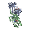
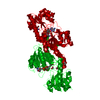
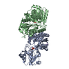
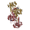
The asymmetric unit has two molecules, A and B. The A molecule is bound to NADP, whereas there is no density for the NADP in the B molecule. The A molecule is also much better ordered than the B molecule in the small domain.
-
Components
#1: Protein
FORMATEDEHYDROGENASE
Mass: 34026.176 Da / Num. of mol.: 2 Source method: isolated from a genetically manipulated source Details: FULL LENGTH PROTEIN WITH C-TERM HIS TAG (HHHHHH). HIS TAG WAS NOT SEEN IN THE DENSITY. Source: (gene. exp.) Pyrobaculum aerophilum (archaea) / Description: hyperthermophilic archeabacterium / Production host: Escherichia coli (E. coli) / References: UniProt: Q8ZXP5
Mass: 18.015 Da / Num. of mol.: 3 / Source method: isolated from a natural source / Formula: H2O
Has protein modification
Y
-
Experimental details
-
Experiment
Experiment
Method: X-RAY DIFFRACTION / Number of used crystals: 1
-
Sample preparation
Crystal
Density Matthews: 4.05 Å3/Da / Density % sol: 70 %
Crystal grow
Temperature: 293 K / Method: vapor diffusion, hanging drop / pH: 5.8 Details: about 3M formate at pH 5.8, VAPOR DIFFUSION, HANGING DROP, temperature 293K
Resolution: 2.8→50 Å / Cross valid method: FREE R / σ(F): 0 / σ(I): 0 / Stereochemistry target values: Engh & Huber / Details: The bulk solvent was used as implemented in CNS.
Rfactor
Num. reflection
% reflection
Rfree
0.32
1417
5 %
Rwork
0.274
-
-
all
0.274
28370
-
obs
0.274
28352
99.9 %
Displacement parameters
Biso mean: 59.2 Å2
Refinement step
Cycle: LAST / Resolution: 2.8→50 Å
Protein
Nucleic acid
Ligand
Solvent
Total
Num. atoms
4740
0
48
3
4791
Refine LS restraints
Refine-ID
Type
Dev ideal
X-RAY DIFFRACTION
x_bond_d
0.008
X-RAY DIFFRACTION
x_angle_deg
1.7
+
About Yorodumi
-
News
-
Feb 9, 2022. New format data for meta-information of EMDB entries
New format data for meta-information of EMDB entries
Version 3 of the EMDB header file is now the official format.
The previous official version 1.9 will be removed from the archive.
In the structure databanks used in Yorodumi, some data are registered as the other names, "COVID-19 virus" and "2019-nCoV". Here are the details of the virus and the list of structure data.
Jan 31, 2019. EMDB accession codes are about to change! (news from PDBe EMDB page)
EMDB accession codes are about to change! (news from PDBe EMDB page)
The allocation of 4 digits for EMDB accession codes will soon come to an end. Whilst these codes will remain in use, new EMDB accession codes will include an additional digit and will expand incrementally as the available range of codes is exhausted. The current 4-digit format prefixed with “EMD-” (i.e. EMD-XXXX) will advance to a 5-digit format (i.e. EMD-XXXXX), and so on. It is currently estimated that the 4-digit codes will be depleted around Spring 2019, at which point the 5-digit format will come into force.
The EM Navigator/Yorodumi systems omit the EMD- prefix.
Related info.:Q: What is EMD? / ID/Accession-code notation in Yorodumi/EM Navigator
Yorodumi is a browser for structure data from EMDB, PDB, SASBDB, etc.
This page is also the successor to EM Navigator detail page, and also detail information page/front-end page for Omokage search.
The word "yorodu" (or yorozu) is an old Japanese word meaning "ten thousand". "mi" (miru) is to see.
Related info.:EMDB / PDB / SASBDB / Comparison of 3 databanks / Yorodumi Search / Aug 31, 2016. New EM Navigator & Yorodumi / Yorodumi Papers / Jmol/JSmol / Function and homology information / Changes in new EM Navigator and Yorodumi
 Movie
Movie Controller

 Yorodumi
 Open data
 Basic information
 Components
 Keywords
 Function and homology information
 Pyrobaculum aerophilum (archaea)
 X-RAY DIFFRACTION /
  Authors
 Citation
 Structure visualization
 Downloads & links
 Other downloads
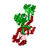


 PDBj
PDBj

 Assembly


 Mass: 745.421 Da / Num. of mol.: 1 / Source method: obtained synthetically / Formula: C21H30N7O17P3
 Mass: 18.015 Da / Num. of mol.: 3 / Source method: isolated from a natural source / Formula: H2O
 Sample preparation
 / Beamline: X8C / Wavelength: 0.98
  Processing