Signaling by TCF7L2 mutants / Repression of WNT target genes / synaptic vesicle clustering / presynaptic active zone cytoplasmic component / Oxidoreductases; Acting on the CH-OH group of donors; With NAD+ or NADP+ as acceptor / oxidoreductase activity, acting on the CH-OH group of donors, NAD or NADP as acceptor / lncRNA binding / white fat cell differentiation / Notch signaling pathway / synaptic vesicle endocytosis ...Signaling by TCF7L2 mutants / Repression of WNT target genes / synaptic vesicle clustering / presynaptic active zone cytoplasmic component / Oxidoreductases; Acting on the CH-OH group of donors; With NAD+ or NADP+ as acceptor / oxidoreductase activity, acting on the CH-OH group of donors, NAD or NADP as acceptor / lncRNA binding / white fat cell differentiation / Notch signaling pathway / synaptic vesicle endocytosis / transcription repressor complex / SUMOylation of transcription cofactors / viral genome replication / transcription corepressor binding / transcription coregulator binding / Deactivation of the beta-catenin transactivating complex / Negative Regulation of CDH1 Gene Transcription / GABA-ergic synapse / NAD binding / transcription corepressor activity / DNA-binding transcription factor binding / RNA polymerase II-specific DNA-binding transcription factor binding / protein phosphorylation / transcription coactivator activity / regulation of cell cycle / negative regulation of cell population proliferation / protein domain specific binding / negative regulation of DNA-templated transcription / chromatin binding / regulation of transcription by RNA polymerase II / glutamatergic synapse / negative regulation of transcription by RNA polymerase II / nucleoplasm / identical protein binding / nucleus Similarity search - Function
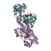
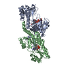
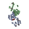
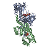
C-terminal binding protein / : / D-isomer specific 2-hydroxyacid dehydrogenases NAD-binding signature. / D-isomer specific 2-hydroxyacid dehydrogenases signature 3. / D-isomer specific 2-hydroxyacid dehydrogenase, NAD-binding domain conserved site 1 / D-isomer specific 2-hydroxyacid dehydrogenase, NAD-binding domain conserved site / D-isomer specific 2-hydroxyacid dehydrogenase, catalytic domain / D-isomer specific 2-hydroxyacid dehydrogenase, catalytic domain / D-isomer specific 2-hydroxyacid dehydrogenase, NAD-binding domain / D-isomer specific 2-hydroxyacid dehydrogenase, NAD binding domain ...C-terminal binding protein / : / D-isomer specific 2-hydroxyacid dehydrogenases NAD-binding signature. / D-isomer specific 2-hydroxyacid dehydrogenases signature 3. / D-isomer specific 2-hydroxyacid dehydrogenase, NAD-binding domain conserved site 1 / D-isomer specific 2-hydroxyacid dehydrogenase, NAD-binding domain conserved site / D-isomer specific 2-hydroxyacid dehydrogenase, catalytic domain / D-isomer specific 2-hydroxyacid dehydrogenase, catalytic domain / D-isomer specific 2-hydroxyacid dehydrogenase, NAD-binding domain / D-isomer specific 2-hydroxyacid dehydrogenase, NAD binding domain / NAD(P)-binding Rossmann-like Domain / NAD(P)-binding domain superfamily / Rossmann fold / 3-Layer(aba) Sandwich / Alpha Beta Similarity search - Domain/homology
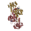
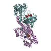
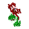
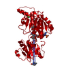
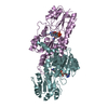
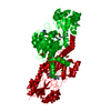
Mass: 38139.469 Da / Num. of mol.: 1 / Fragment: NAD nucleotide binding residues 28-353 Source method: isolated from a genetically manipulated source Source: (gene. exp.) Homo sapiens (human) / Gene: CTBP1, CTBP / Plasmid: pet28a / Production host: Escherichia coli (E. coli) / Strain (production host): BL21-DE3 / Variant (production host): RIL+ References: UniProt: Q13363, Oxidoreductases; Acting on the CH-OH group of donors; With NAD+ or NADP+ as acceptor
In the structure databanks used in Yorodumi, some data are registered as the other names, "COVID-19 virus" and "2019-nCoV". Here are the details of the virus and the list of structure data.
Jan 31, 2019. EMDB accession codes are about to change! (news from PDBe EMDB page)
EMDB accession codes are about to change! (news from PDBe EMDB page)
The allocation of 4 digits for EMDB accession codes will soon come to an end. Whilst these codes will remain in use, new EMDB accession codes will include an additional digit and will expand incrementally as the available range of codes is exhausted. The current 4-digit format prefixed with “EMD-” (i.e. EMD-XXXX) will advance to a 5-digit format (i.e. EMD-XXXXX), and so on. It is currently estimated that the 4-digit codes will be depleted around Spring 2019, at which point the 5-digit format will come into force.
The EM Navigator/Yorodumi systems omit the EMD- prefix.
Related info.:Q: What is EMD? / ID/Accession-code notation in Yorodumi/EM Navigator
Yorodumi is a browser for structure data from EMDB, PDB, SASBDB, etc.
This page is also the successor to EM Navigator detail page, and also detail information page/front-end page for Omokage search.
The word "yorodu" (or yorozu) is an old Japanese word meaning "ten thousand". "mi" (miru) is to see.
Related info.:EMDB / PDB / SASBDB / Comparison of 3 databanks / Yorodumi Search / Aug 31, 2016. New EM Navigator & Yorodumi / Yorodumi Papers / Jmol/JSmol / Function and homology information / Changes in new EM Navigator and Yorodumi
 Movie
Movie Controller


 Open data
 Basic information
 Components
 Keywords
 Function and homology information
 Homo sapiens (human)
 X-RAY DIFFRACTION /
  Authors
 Citation
 Structure visualization
 Downloads & links
 Other downloads
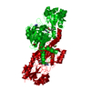


 PDBj
PDBj


 Assembly
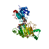


 Mass: 665.441 Da / Num. of mol.: 1 / Source method: obtained synthetically / Formula: C21H29N7O14P2
 Mass: 179.173 Da / Num. of mol.: 1 / Source method: obtained synthetically / Formula: C9H9NO3
 Mass: 40.078 Da / Num. of mol.: 1 / Source method: obtained synthetically / Formula: Ca
 Mass: 46.025 Da / Num. of mol.: 1 / Source method: obtained synthetically / Formula: CH2O2
 Sample preparation
 Processing