[English] 日本語
Yorodumi
- PDB-2ome: Crystal structure of human CTBP2 dehydrogenase complexed with NAD(H) -

+
Open data


ID or keywords:

Loading...

-
Basic information

Entry
Database: PDB / ID: 2ome
TitleCrystal structure of human CTBP2 dehydrogenase complexed with NAD(H)
ComponentsC-terminal-binding protein 2
KeywordsOXIDOREDUCTASE / C-TERMINAL BINDING PROTEIN / CTBP2 / DEHYDROGENASE / STRUCTURAL GENOMICS CONSORTIUM / SGC
Function / homology
Function and homology information


positive regulation of retinoic acid receptor signaling pathway / structural constituent of presynaptic active zone / Signaling by TCF7L2 mutants / Repression of WNT target genes / synaptic vesicle docking / photoreceptor ribbon synapse / presynaptic active zone cytoplasmic component / nuclear retinoic acid receptor binding / presynaptic cytosol / Sensory processing of sound by inner hair cells of the cochlea ...positive regulation of retinoic acid receptor signaling pathway / structural constituent of presynaptic active zone / Signaling by TCF7L2 mutants / Repression of WNT target genes / synaptic vesicle docking / photoreceptor ribbon synapse / presynaptic active zone cytoplasmic component / nuclear retinoic acid receptor binding / presynaptic cytosol / Sensory processing of sound by inner hair cells of the cochlea / oxidoreductase activity, acting on the CH-OH group of donors, NAD or NADP as acceptor / white fat cell differentiation / GABA-ergic synapse / transcription repressor complex / cellular response to leukemia inhibitory factor / viral genome replication / transcription corepressor binding / transcription coregulator binding / transcription corepressor activity / NAD binding / DNA-binding transcription factor binding / transcription coactivator activity / negative regulation of cell population proliferation / negative regulation of DNA-templated transcription / glutamatergic synapse / chromatin binding / protein-containing complex binding / regulation of transcription by RNA polymerase II / protein kinase binding / negative regulation of transcription by RNA polymerase II / positive regulation of transcription by RNA polymerase II / identical protein binding / nucleus
Similarity search - Function
C-terminal binding protein / D-isomer specific 2-hydroxyacid dehydrogenases signature 3. / D-isomer specific 2-hydroxyacid dehydrogenase, NAD-binding domain conserved site / D-isomer specific 2-hydroxyacid dehydrogenase, catalytic domain / D-isomer specific 2-hydroxyacid dehydrogenase, catalytic domain / D-isomer specific 2-hydroxyacid dehydrogenase, NAD-binding domain / D-isomer specific 2-hydroxyacid dehydrogenase, NAD binding domain / NAD(P)-binding Rossmann-like Domain / NAD(P)-binding domain superfamily / Rossmann fold ...C-terminal binding protein / D-isomer specific 2-hydroxyacid dehydrogenases signature 3. / D-isomer specific 2-hydroxyacid dehydrogenase, NAD-binding domain conserved site / D-isomer specific 2-hydroxyacid dehydrogenase, catalytic domain / D-isomer specific 2-hydroxyacid dehydrogenase, catalytic domain / D-isomer specific 2-hydroxyacid dehydrogenase, NAD-binding domain / D-isomer specific 2-hydroxyacid dehydrogenase, NAD binding domain / NAD(P)-binding Rossmann-like Domain / NAD(P)-binding domain superfamily / Rossmann fold / 3-Layer(aba) Sandwich / Alpha Beta
Similarity search - Domain/homology
NICOTINAMIDE-ADENINE-DINUCLEOTIDE / C-terminal-binding protein 2
Similarity search - Component
Biological speciesHomo sapiens (human)
MethodX-RAY DIFFRACTION / SYNCHROTRON / MOLECULAR REPLACEMENT / Resolution: 2.8 Å
AuthorsPilka, E.S. / Guo, K. / Rojkova, A. / Debreczeni, J.E. / Kavanagh, K.L. / von Delft, F. / Arrowsmith, C.H. / Weigelt, J. / Edwards, A. / Sundstrom, M. ...Pilka, E.S. / Guo, K. / Rojkova, A. / Debreczeni, J.E. / Kavanagh, K.L. / von Delft, F. / Arrowsmith, C.H. / Weigelt, J. / Edwards, A. / Sundstrom, M. / Oppermann, U. / Structural Genomics Consortium (SGC)
CitationJournal: To be Published
Title: Crystal structure of human CTBP2 dehydrogenase complexed with NAD(H)
Authors: Pilka, E.S. / Guo, K. / Rojkova, A. / Debreczeni, J.E. / Kavanagh, K.L. / von Delft, F. / Arrowsmith, C.H. / Weigelt, J. / Edwards, A. / Sundstrom, M. / Oppermann, U.
History
DepositionJan 22, 2007Deposition site: RCSB / Processing site: RCSB
Revision 1.0Feb 6, 2007Provider: repository / Type: Initial release
Revision 1.1May 1, 2008Group: Version format compliance
Revision 1.2Jul 13, 2011Group: Advisory / Version format compliance
Revision 1.3Oct 18, 2017Group: Refinement description / Category: software / Item: _software.name
Revision 1.4Aug 30, 2023Group: Data collection / Database references ...Data collection / Database references / Derived calculations / Refinement description
Category: chem_comp_atom / chem_comp_bond ...chem_comp_atom / chem_comp_bond / database_2 / pdbx_initial_refinement_model / struct_ncs_dom_lim / struct_ref_seq_dif / struct_site
Item: _database_2.pdbx_DOI / _database_2.pdbx_database_accession ..._database_2.pdbx_DOI / _database_2.pdbx_database_accession / _struct_ncs_dom_lim.beg_auth_comp_id / _struct_ncs_dom_lim.end_auth_comp_id / _struct_ref_seq_dif.details / _struct_site.pdbx_auth_asym_id / _struct_site.pdbx_auth_comp_id / _struct_site.pdbx_auth_seq_id

-
Structure visualization

Structure viewerMolecule:
MolmilJmol/JSmol

Downloads & links

-
Assembly

Deposited unit
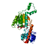
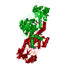
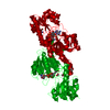
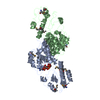
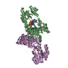
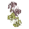
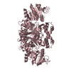
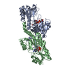
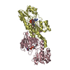
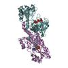
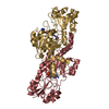
A: C-terminal-binding protein 2
B: C-terminal-binding protein 2
C: C-terminal-binding protein 2
D: C-terminal-binding protein 2
E: C-terminal-binding protein 2
F: C-terminal-binding protein 2
G: C-terminal-binding protein 2
H: C-terminal-binding protein 2
hetero molecules


Theoretical massNumber of molelcules
Total (without water)303,50316
Polymers298,1968
Non-polymers5,3078
Water3,315184
1
A: C-terminal-binding protein 2
B: C-terminal-binding protein 2
hetero molecules


Theoretical massNumber of molelcules
Total (without water)75,8764
Polymers74,5492
Non-polymers1,3272
Water362
TypeNameSymmetry operationNumber
identity operation1_555x,y,z1
Buried area8470 Å2
ΔGint-37 kcal/mol
Surface area25790 Å2
MethodPISA
2
C: C-terminal-binding protein 2
D: C-terminal-binding protein 2
hetero molecules


Theoretical massNumber of molelcules
Total (without water)75,8764
Polymers74,5492
Non-polymers1,3272
Water362
TypeNameSymmetry operationNumber
identity operation1_555x,y,z1
Buried area8460 Å2
ΔGint-38 kcal/mol
Surface area25990 Å2
MethodPISA
3
E: C-terminal-binding protein 2
F: C-terminal-binding protein 2
hetero molecules


Theoretical massNumber of molelcules
Total (without water)75,8764
Polymers74,5492
Non-polymers1,3272
Water362
TypeNameSymmetry operationNumber
identity operation1_555x,y,z1
Buried area8510 Å2
ΔGint-39 kcal/mol
Surface area25700 Å2
MethodPISA
4
G: C-terminal-binding protein 2
H: C-terminal-binding protein 2
hetero molecules


Theoretical massNumber of molelcules
Total (without water)75,8764
Polymers74,5492
Non-polymers1,3272
Water362
TypeNameSymmetry operationNumber
identity operation1_555x,y,z1
Buried area8460 Å2
ΔGint-40 kcal/mol
Surface area25540 Å2
MethodPISA
Unit cell
Length a, b, c (Å)87.754, 141.574, 138.215
Angle α, β, γ (deg.)90.00, 98.93, 90.00
Int Tables number4
Space group name H-MP1211
Noncrystallographic symmetry (NCS)NCS domain:
IDEns-IDDetails
11A
21B
31C
41D
51E
61F
71G
81H

NCS domain segments:

Component-ID: 1 / Ens-ID: 1 / Beg auth comp-ID: PRO / Beg label comp-ID: PRO / End auth comp-ID: PHE / End label comp-ID: PHE / Refine code: 3 / Auth seq-ID: 34 - 362 / Label seq-ID: 6 - 334

Dom-IDAuth asym-IDLabel asym-ID
1AA
2BB
3CC
4DD
5EE
6FF
7GG
8HH

-
Components

#1: Protein
C-terminal-binding protein 2 / CtBP2


Mass: 37274.477 Da / Num. of mol.: 8 / Fragment: Residues 31-364
Source method: isolated from a genetically manipulated source
Source: (gene. exp.) Homo sapiens (human) / Gene: CTBP2 / Plasmid: pNIC28-Bsa4 / Species (production host): Escherichia coli / Production host: Escherichia coli BL21(DE3) (bacteria) / Strain (production host): BL21(DE3) / References: UniProt: P56545
#2: Chemical
ChemComp-NAD / NICOTINAMIDE-ADENINE-DINUCLEOTIDE / Nicotinamide adenine dinucleotide


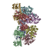
Mass: 663.425 Da / Num. of mol.: 8 / Source method: obtained synthetically / Formula: C21H27N7O14P2 / Comment: NAD*YM
#3: Water ChemComp-HOH / water / Water


Mass: 18.015 Da / Num. of mol.: 184 / Source method: isolated from a natural source / Formula: H2O

-
Experimental details

-
Experiment

ExperimentMethod: X-RAY DIFFRACTION / Number of used crystals: 1

-
Sample preparation

CrystalDensity Matthews: 2.84 Å3/Da / Density % sol: 56.74 %
Crystal growTemperature: 277 K / Method: vapor diffusion, sitting drop / pH: 6.5
Details: 0.2M KSCN, 0.1M BIS-TRIS PROPANE, 20% PEG3350, 10% ETHYLENE GLYCOL, pH 6.5, VAPOR DIFFUSION, SITTING DROP, temperature 277K

-
Data collection

DiffractionMean temperature: 100 K
Diffraction sourceSource: SYNCHROTRON / Site: SLS / Beamline: X10SA / Wavelength: 0.99991 Å
DetectorType: MARMOSAIC 225 mm CCD / Detector: CCD / Date: Dec 21, 2006
RadiationProtocol: SINGLE WAVELENGTH / Monochromatic (M) / Laue (L): M / Scattering type: x-ray
Radiation wavelengthWavelength: 0.99991 Å / Relative weight: 1
ReflectionResolution: 2.77→43.31 Å / Num. obs: 84796 / % possible obs: 100 % / Observed criterion σ(I): 2 / Redundancy: 6.1 % / Rmerge(I) obs: 0.148 / Rsym value: 0.162 / Net I/σ(I): 11.6
Reflection shellResolution: 2.77→2.92 Å / Redundancy: 5.9 % / Rmerge(I) obs: 0.81 / Mean I/σ(I) obs: 2 / Rsym value: 0.89 / % possible all: 100

-
Processing

Software
NameVersionClassification
REFMAC5.2.0019refinement
MAR345345DTBdata collection
MOSFLMdata reduction
CCP4(SCALA)data scaling
PHASERphasing
RefinementMethod to determine structure: MOLECULAR REPLACEMENT
Starting model: PDB entries 1MX3, 1HL3
Resolution: 2.8→43.31 Å / Cor.coef. Fo:Fc: 0.933 / Cor.coef. Fo:Fc free: 0.9 / SU B: 30.465 / SU ML: 0.3 / TLS residual ADP flag: LIKELY RESIDUAL / Cross valid method: THROUGHOUT / σ(F): 2 / ESU R Free: 0.378 / Stereochemistry target values: MAXIMUM LIKELIHOOD / Details: HYDROGENS HAVE BEEN ADDED IN THE RIDING POSITIONS
RfactorNum. reflection% reflectionSelection details
Rfree0.25736 4104 5 %RANDOM
Rwork0.20733 ---
obs0.2098 78004 99.98 %-
all-84796 --
Solvent computationIon probe radii: 0.8 Å / Shrinkage radii: 0.8 Å / VDW probe radii: 1.4 Å / Solvent model: MASK
Displacement parametersBiso mean: 40.724 Å2
Baniso -1Baniso -2Baniso -3
1-1.63 Å20 Å2-0.55 Å2
2---4.83 Å20 Å2
3---3.03 Å2
Refinement stepCycle: LAST / Resolution: 2.8→43.31 Å
ProteinNucleic acidLigandSolventTotal
Num. atoms20223 0 352 184 20759
Refine LS restraints
Refine-IDTypeDev idealDev ideal targetNumber
X-RAY DIFFRACTIONr_bond_refined_d0.0130.02120977
X-RAY DIFFRACTIONr_bond_other_d0.0020.0213837
X-RAY DIFFRACTIONr_angle_refined_deg1.4581.97328515
X-RAY DIFFRACTIONr_angle_other_deg1.116333574
X-RAY DIFFRACTIONr_dihedral_angle_1_deg6.34852635
X-RAY DIFFRACTIONr_dihedral_angle_2_deg35.74423.508972
X-RAY DIFFRACTIONr_dihedral_angle_3_deg17.854153379
X-RAY DIFFRACTIONr_dihedral_angle_4_deg17.54215188
X-RAY DIFFRACTIONr_chiral_restr0.0750.23283
X-RAY DIFFRACTIONr_gen_planes_refined0.0040.0223591
X-RAY DIFFRACTIONr_gen_planes_other0.0020.024305
X-RAY DIFFRACTIONr_nbd_refined0.2110.24159
X-RAY DIFFRACTIONr_nbd_other0.1950.214266
X-RAY DIFFRACTIONr_nbtor_refined0.1840.210078
X-RAY DIFFRACTIONr_nbtor_other0.0870.211775
X-RAY DIFFRACTIONr_xyhbond_nbd_refined0.1490.2404
X-RAY DIFFRACTIONr_xyhbond_nbd_other0.0230.21
X-RAY DIFFRACTIONr_metal_ion_refined
X-RAY DIFFRACTIONr_metal_ion_other
X-RAY DIFFRACTIONr_symmetry_vdw_refined0.1810.210
X-RAY DIFFRACTIONr_symmetry_vdw_other0.1780.218
X-RAY DIFFRACTIONr_symmetry_hbond_refined0.270.22
X-RAY DIFFRACTIONr_symmetry_hbond_other
X-RAY DIFFRACTIONr_symmetry_metal_ion_refined
X-RAY DIFFRACTIONr_symmetry_metal_ion_other
X-RAY DIFFRACTIONr_mcbond_it0.3491.513575
X-RAY DIFFRACTIONr_mcbond_other0.1281.55390
X-RAY DIFFRACTIONr_mcangle_it0.548220924
X-RAY DIFFRACTIONr_scbond_it1.25338470
X-RAY DIFFRACTIONr_scangle_it2.0144.57591
X-RAY DIFFRACTIONr_rigid_bond_restr
X-RAY DIFFRACTIONr_sphericity_free
X-RAY DIFFRACTIONr_sphericity_bonded
Refine LS restraints NCS

Ens-ID: 1 / Refine-ID: X-RAY DIFFRACTION

Dom-IDAuth asym-IDNumberTypeRms dev position (Å)Weight position
1A1930tight positional0.040.05
2B1930tight positional0.040.05
3C1930tight positional0.040.05
4D1930tight positional0.040.05
5E1930tight positional0.040.05
6F1930tight positional0.040.05
7G1930tight positional0.030.05
8H1930tight positional0.040.05
1A2050loose positional0.45
2B2050loose positional0.375
3C2050loose positional0.395
4D2050loose positional0.455
5E2050loose positional0.515
6F2050loose positional0.45
7G2050loose positional0.475
8H2050loose positional0.425
1A1930tight thermal0.070.5
2B1930tight thermal0.070.5
3C1930tight thermal0.080.5
4D1930tight thermal0.070.5
5E1930tight thermal0.060.5
6F1930tight thermal0.070.5
7G1930tight thermal0.060.5
8H1930tight thermal0.070.5
1A2050loose thermal1.2310
2B2050loose thermal1.1610
3C2050loose thermal1.1110
4D2050loose thermal1.0110
5E2050loose thermal1.1410
6F2050loose thermal1.0810
7G2050loose thermal1.0510
8H2050loose thermal1.0610
LS refinement shellResolution: 2.8→2.872 Å / Total num. of bins used: 20
RfactorNum. reflection% reflection
Rfree0.387 298 -
Rwork0.361 5765 -
obs--100 %
Refinement TLS params.

Method: refined / Refine-ID: X-RAY DIFFRACTION

IDL112)L122)L132)L222)L232)L332)S11 (Å °)S12 (Å °)S13 (Å °)S21 (Å °)S22 (Å °)S23 (Å °)S31 (Å °)S32 (Å °)S33 (Å °)T112)T122)T132)T222)T232)T332)Origin x (Å)Origin y (Å)Origin z (Å)
11.22180.4041-0.10773.4584-0.60241.64920.12840.1556-0.3536-0.4701-0.10880.21220.28520.1155-0.0196-0.17010.0778-0.2226-0.1656-0.1082-0.094215.525-3.54715.2843
21.5598-0.5827-0.53662.76680.59262.14570.2530.22050.2951-0.4782-0.0566-0.466-0.39310.3094-0.1964-0.1784-0.00760.0327-0.058-0.0437-0.162632.186527.72676.9872
31.53880.5130.44243.30650.5641.4577-0.032-0.0899-0.05850.3652-0.10720.4182-0.2301-0.04190.1392-0.13430.068-0.0124-0.2394-0.0098-0.270110.237438.450435.2989
41.48040.34210.2571.80880.52161.5408-0.0559-0.2185-0.3540.2975-0.14230.85190.1522-0.41370.1982-0.1511-0.00730.1554-0.1283-0.05690.3615-7.62067.526832.1255
51.63820.44880.312.65960.4011.31040.0973-0.40860.39560.8789-0.22551.2529-0.0532-0.39910.12820.134-0.02960.4479-0.0617-0.08730.46693.0779-28.309453.5182
61.04430.56530.23273.54481.14322.10790.2259-0.0160.13130.282-0.11820.7620.1945-0.1163-0.1077-0.1371-0.00820.0471-0.21910.0708-0.052411.8168-59.161138.2784
72.62750.8143-0.07853.44890.31941.889-0.14750.0704-0.83630.16220.047-0.86120.43680.54150.1004-0.11850.11630.09340.03540.03040.058447.472-47.992938.4223
81.3617-0.5220.40364.6161-0.2361.6389-0.01170.0110.18970.6038-0.1399-0.2861-0.23280.22130.15160.0124-0.0605-0.0434-0.10730.0528-0.322438.3738-16.975852.5326
Refinement TLS group
IDRefine-IDRefine TLS-IDAuth asym-IDLabel asym-IDAuth seq-IDLabel seq-ID
1X-RAY DIFFRACTION1AA33 - 3625 - 334
2X-RAY DIFFRACTION2BB33 - 3625 - 334
3X-RAY DIFFRACTION3CC33 - 3625 - 334
4X-RAY DIFFRACTION4DD33 - 3625 - 334
5X-RAY DIFFRACTION5EE33 - 3625 - 334
6X-RAY DIFFRACTION6FF33 - 3625 - 334
7X-RAY DIFFRACTION7GG33 - 3625 - 334
8X-RAY DIFFRACTION8HH33 - 3625 - 334

+
About Yorodumi

-
News

-
Feb 9, 2022. New format data for meta-information of EMDB entries

New format data for meta-information of EMDB entries

  • Version 3 of the EMDB header file is now the official format.
  • The previous official version 1.9 will be removed from the archive.

Related info.:EMDB header

External links:wwPDB to switch to version 3 of the EMDB data model

-
Aug 12, 2020. Covid-19 info

Covid-19 info

URL: https://pdbj.org/emnavi/covid19.php

New page: Covid-19 featured information page in EM Navigator.

Related info.:Covid-19 info / Mar 5, 2020. Novel coronavirus structure data

+
Mar 5, 2020. Novel coronavirus structure data

Novel coronavirus structure data

Related info.:Yorodumi Speices / Aug 12, 2020. Covid-19 info

External links:COVID-19 featured content - PDBj / Molecule of the Month (242):Coronavirus Proteases

+
Jan 31, 2019. EMDB accession codes are about to change! (news from PDBe EMDB page)

EMDB accession codes are about to change! (news from PDBe EMDB page)

  • The allocation of 4 digits for EMDB accession codes will soon come to an end. Whilst these codes will remain in use, new EMDB accession codes will include an additional digit and will expand incrementally as the available range of codes is exhausted. The current 4-digit format prefixed with “EMD-” (i.e. EMD-XXXX) will advance to a 5-digit format (i.e. EMD-XXXXX), and so on. It is currently estimated that the 4-digit codes will be depleted around Spring 2019, at which point the 5-digit format will come into force.
  • The EM Navigator/Yorodumi systems omit the EMD- prefix.

Related info.:Q: What is EMD? / ID/Accession-code notation in Yorodumi/EM Navigator

External links:EMDB Accession Codes are Changing Soon! / Contact to PDBj

+
Jul 12, 2017. Major update of PDB

Major update of PDB

  • wwPDB released updated PDB data conforming to the new PDBx/mmCIF dictionary.
  • This is a major update changing the version number from 4 to 5, and with Remediation, in which all the entries are updated.
  • In this update, many items about electron microscopy experimental information are reorganized (e.g. em_software).
  • Now, EM Navigator and Yorodumi are based on the updated data.

External links:wwPDB Remediation / Enriched Model Files Conforming to OneDep Data Standards Now Available in the PDB FTP Archive

-
Yorodumi

Thousand views of thousand structures

  • Yorodumi is a browser for structure data from EMDB, PDB, SASBDB, etc.
  • This page is also the successor to EM Navigator detail page, and also detail information page/front-end page for Omokage search.
  • The word "yorodu" (or yorozu) is an old Japanese word meaning "ten thousand". "mi" (miru) is to see.

Related info.:EMDB / PDB / SASBDB / Comparison of 3 databanks / Yorodumi Search / Aug 31, 2016. New EM Navigator & Yorodumi / Yorodumi Papers / Jmol/JSmol / Function and homology information / Changes in new EM Navigator and Yorodumi

Read more