 Movie
Movie Controller

[English] 日本語
 Yorodumi
- PDB-1hku: CtBP/BARS: a dual-function protein involved in transcription core... -
+ Open data

- Basic information
| Entry | Database: PDB / ID: 1hku | ||||||
|---|---|---|---|---|---|---|---|
| Title | CtBP/BARS: a dual-function protein involved in transcription corepression and Golgi membrane fission | ||||||
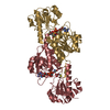
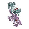
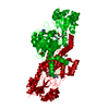
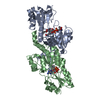
 Components Components | C-TERMINAL BINDING PROTEIN 3 | ||||||
 Keywords Keywords | TRANSCRIPTION / TRANSCRIPTION CO-REPRESSOR / TRANSCRIPTION CO-REPRESSION / ACYLTRANSFERASE / BREFELDIN A / NAD / GOLGI MEMBRANE / ACYL-COA | ||||||
| Function / homology |  Function and homology information Function and homology informationpresynapse to nucleus signaling pathway / SUMOylation of transcription cofactors / Repression of WNT target genes / Negative Regulation of CDH1 Gene Transcription / Deactivation of the beta-catenin transactivating complex / extrinsic component of presynaptic endocytic zone membrane / synaptic vesicle clustering / presynaptic active zone cytoplasmic component / Oxidoreductases; Acting on the CH-OH group of donors; With NAD+ or NADP+ as acceptor / oxidoreductase activity, acting on the CH-OH group of donors, NAD or NADP as acceptor ...presynapse to nucleus signaling pathway / SUMOylation of transcription cofactors / Repression of WNT target genes / Negative Regulation of CDH1 Gene Transcription / Deactivation of the beta-catenin transactivating complex / extrinsic component of presynaptic endocytic zone membrane / synaptic vesicle clustering / presynaptic active zone cytoplasmic component / Oxidoreductases; Acting on the CH-OH group of donors; With NAD+ or NADP+ as acceptor / oxidoreductase activity, acting on the CH-OH group of donors, NAD or NADP as acceptor / acyltransferase activity / lncRNA binding / white fat cell differentiation / Notch signaling pathway / synaptic vesicle endocytosis / transcription repressor complex / transcription corepressor binding / transcription coregulator binding / PDZ domain binding / GABA-ergic synapse / NAD binding / transcription corepressor activity / DNA-binding transcription factor binding / membrane fusion / RNA polymerase II-specific DNA-binding transcription factor binding / transcription coactivator activity / regulation of cell cycle / neuron projection / protein domain specific binding / negative regulation of DNA-templated transcription / chromatin binding / regulation of transcription by RNA polymerase II / glutamatergic synapse / negative regulation of transcription by RNA polymerase II / protein homodimerization activity / nucleoplasm / identical protein binding / nucleus / cytoplasm Similarity search - Function | ||||||
| Biological species |  | ||||||
| Method |  X-RAY DIFFRACTION / SYNCHROTRON / MAD / Resolution: 2.3 Å X-RAY DIFFRACTION / SYNCHROTRON / MAD / Resolution: 2.3 Å | ||||||
 Authors Authors | Nardini, M. / Spano, S. / Cericola, C. / Pesce, A. / Massaro, A. / Millo, E. / Luini, A. / Corda, D. / Bolognesi, M. | ||||||
 Citation Citation | Journal: Embo J. / Year: 2003 Title: Ctbp/Bars: A Dual-Function Protein Involved in Transcription Co-Repression and Golgi Membrane Fission Authors: Nardini, M. / Spano, S. / Cericola, C. / Pesce, A. / Massaro, A. / Millo, E. / Luini, A. / Corda, D. / Bolognesi, M. #1: Journal: Acta Crystallogr.,Sect.D / Year: 2002 Title: Crystallization and Preliminary X-Ray Diffraction Analysis of Brefeldin A-Adp Ribosylated Substrate (Bars) Authors: Nardini, M. / Spano, S. / Cericola, C. / Pesce, A. / Damonte, G. / Luini, A. / Corda, D. / Bolognesi, M. | ||||||
| History |
|
- Structure visualization
| Structure viewer | Molecule: MolmilJmol/JSmol |
|---|
- Downloads & links
-Download
| PDBx/mmCIF format | 1hku.cif.gz | 82.6 KB | Display | PDBx/mmCIF format |
|---|---|---|---|---|
| PDB format | pdb1hku.ent.gz | 60.7 KB | Display | PDB format |
| PDBx/mmJSON format | 1hku.json.gz | Tree view | PDBx/mmJSON format | |
| Others |  Other downloads Other downloads |
-Validation report
| Arichive directory | https://data.pdbj.org/pub/pdb/validation_reports/hk/1hkuftp://data.pdbj.org/pub/pdb/validation_reports/hk/1hku | HTTPS FTP |
|---|
-Related structure data


-Links
 PDBj
PDBj- Assembly
| Deposited unit | 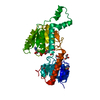
| ||||||||
|---|---|---|---|---|---|---|---|---|---|
| 1 | 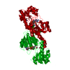
| ||||||||
| Unit cell |
|
-Components
| #1: Protein | Mass: 39525.133 Da / Num. of mol.: 1 / Fragment: RESIDUES 1-350 Source method: isolated from a genetically manipulated source Source: (gene. exp.)  | ||||||||
|---|---|---|---|---|---|---|---|---|---|
| #2: Chemical | ChemComp-NAD /   Mass: 663.425 Da / Num. of mol.: 1 / Source method: obtained synthetically / Formula: C21H27N7O14P2 / Comment: NAD*YM Mass: 663.425 Da / Num. of mol.: 1 / Source method: obtained synthetically / Formula: C21H27N7O14P2 / Comment: NAD*YM | ||||||||
| #3: Chemical | ChemComp-GOL /   Mass: 92.094 Da / Num. of mol.: 1 / Source method: obtained synthetically / Formula: C3H8O3 Mass: 92.094 Da / Num. of mol.: 1 / Source method: obtained synthetically / Formula: C3H8O3 | ||||||||
| #4: Chemical |   Mass: 46.025 Da / Num. of mol.: 2 / Source method: obtained synthetically / Formula: CH2O2 Mass: 46.025 Da / Num. of mol.: 2 / Source method: obtained synthetically / Formula: CH2O2#5: Water | ChemComp-HOH / |  Mass: 18.015 Da / Num. of mol.: 114 / Source method: isolated from a natural source / Formula: H2O Mass: 18.015 Da / Num. of mol.: 114 / Source method: isolated from a natural source / Formula: H2OCompound details | POSSIBLLY NVOLVED IN CONTROLLIN | Has protein modification | Y | Sequence details | HIS-TAG (MGHHHHHH), TRUNCATION | |
-Experimental details
-Experiment
| Experiment | Method: X-RAY DIFFRACTION / Number of used crystals: 1 |
|---|
- Sample preparation
| Crystal | Density Matthews: 2.34 Å3/Da / Density % sol: 47.5 % | ||||||||||||||||||
|---|---|---|---|---|---|---|---|---|---|---|---|---|---|---|---|---|---|---|---|
| Crystal grow | pH: 7.5 / Details: 2.0 M AMMONIUM FORMATE, 100 MM HEPES, PH 7.5. | ||||||||||||||||||
| Crystal grow | *PLUS pH: 7.5 / Method: vapor diffusion, hanging drop | ||||||||||||||||||
| Components of the solutions | *PLUS
|
-Data collection
| Diffraction | Mean temperature: 100 K |
|---|---|
| Diffraction source | Source: SYNCHROTRON / Site: ESRF  / Beamline: ID14-1 / Wavelength: 0.934 / Beamline: ID14-1 / Wavelength: 0.934 |
| Detector | Detector: CCD |
| Radiation | Protocol: SINGLE WAVELENGTH / Monochromatic (M) / Laue (L): M / Scattering type: x-ray |
| Radiation wavelength | Wavelength: 0.934 Å / Relative weight: 1 |
| Reflection | Resolution: 2.3→40 Å / Num. obs: 17070 / % possible obs: 99.3 % / Redundancy: 11 % / Rmerge(I) obs: 0.059 / Net I/σ(I): 25.5 |
| Reflection shell | Resolution: 2.3→2.34 Å / Redundancy: 9 % / Rmerge(I) obs: 0.3 / Mean I/σ(I) obs: 8.7 / % possible all: 100 |
| Reflection | *PLUS Highest resolution: 2.3 Å / Lowest resolution: 40 Å / Redundancy: 11 % / Rmerge(I) obs: 0.059 |
| Reflection shell | *PLUS % possible obs: 100 % / Rmerge(I) obs: 0.302 / Mean I/σ(I) obs: 8.7 |
- Processing
| Software |
| ||||||||||||||||||||||||||||||||||||||||||||||||||||||||||||
|---|---|---|---|---|---|---|---|---|---|---|---|---|---|---|---|---|---|---|---|---|---|---|---|---|---|---|---|---|---|---|---|---|---|---|---|---|---|---|---|---|---|---|---|---|---|---|---|---|---|---|---|---|---|---|---|---|---|---|---|---|---|
| Refinement | Method to determine structure: MAD / Resolution: 2.3→40 Å / σ(F): 2 / Stereochemistry target values: ENGH & HUBER Details: NO DENSITY FOR THE HIS-TAG (MGHHHHHH), AND FOR RESIDUES MET A1-HIS A14 AND THR A346-HIS A350. ATOM OXT OF LEU A345 WAS INTENTIONALLY CONVERTED TO ATOM N OF THR A347 IN ORDER TO COMPLY WITH ...Details: NO DENSITY FOR THE HIS-TAG (MGHHHHHH), AND FOR RESIDUES MET A1-HIS A14 AND THR A346-HIS A350. ATOM OXT OF LEU A345 WAS INTENTIONALLY CONVERTED TO ATOM N OF THR A347 IN ORDER TO COMPLY WITH THE PDB FORMAT. THIS CHANGE MAKES NO DECISION ON THE DIRECTION IN WHICH THE CHAIN BRANCHES.
| ||||||||||||||||||||||||||||||||||||||||||||||||||||||||||||
| Refinement step | Cycle: LAST / Resolution: 2.3→40 Å
| ||||||||||||||||||||||||||||||||||||||||||||||||||||||||||||
| Refine LS restraints |
| ||||||||||||||||||||||||||||||||||||||||||||||||||||||||||||
| Xplor file |
| ||||||||||||||||||||||||||||||||||||||||||||||||||||||||||||
| Refinement | *PLUS Highest resolution: 2.3 Å / % reflection Rfree: 10 % | ||||||||||||||||||||||||||||||||||||||||||||||||||||||||||||
| Solvent computation | *PLUS | ||||||||||||||||||||||||||||||||||||||||||||||||||||||||||||
| Displacement parameters | *PLUS | ||||||||||||||||||||||||||||||||||||||||||||||||||||||||||||
| Refine LS restraints | *PLUS Type: c_angle_deg / Dev ideal: 1.6 |
