[English] 日本語
Yorodumi
- PDB-4lcj: CtBP2 in complex with substrate MTOB -

+
Open data


ID or keywords:

Loading...

-
Basic information

Entry
Database: PDB / ID: 4lcj
TitleCtBP2 in complex with substrate MTOB
ComponentsC-terminal-binding protein 2
KeywordsOXIDOREDUCTASE/OXIDOREDUCTASE SUBSTRATE / Rossmann Fold / Transcriptional Corepressor / D-isomer 2-hydroxyacid dehydrogenase / OXIDOREDUCTASE-OXIDOREDUCTASE SUBSTRATE complex
Function / homology
Function and homology information


positive regulation of retinoic acid receptor signaling pathway / structural constituent of presynaptic active zone / Signaling by TCF7L2 mutants / Repression of WNT target genes / synaptic vesicle docking / photoreceptor ribbon synapse / presynaptic active zone cytoplasmic component / nuclear retinoic acid receptor binding / presynaptic cytosol / Sensory processing of sound by inner hair cells of the cochlea ...positive regulation of retinoic acid receptor signaling pathway / structural constituent of presynaptic active zone / Signaling by TCF7L2 mutants / Repression of WNT target genes / synaptic vesicle docking / photoreceptor ribbon synapse / presynaptic active zone cytoplasmic component / nuclear retinoic acid receptor binding / presynaptic cytosol / Sensory processing of sound by inner hair cells of the cochlea / oxidoreductase activity, acting on the CH-OH group of donors, NAD or NADP as acceptor / white fat cell differentiation / GABA-ergic synapse / transcription repressor complex / cellular response to leukemia inhibitory factor / viral genome replication / transcription corepressor binding / transcription coregulator binding / transcription corepressor activity / NAD binding / DNA-binding transcription factor binding / transcription coactivator activity / negative regulation of cell population proliferation / negative regulation of DNA-templated transcription / glutamatergic synapse / chromatin binding / protein-containing complex binding / regulation of transcription by RNA polymerase II / protein kinase binding / negative regulation of transcription by RNA polymerase II / positive regulation of transcription by RNA polymerase II / identical protein binding / nucleus
Similarity search - Function
C-terminal binding protein / D-isomer specific 2-hydroxyacid dehydrogenases signature 3. / D-isomer specific 2-hydroxyacid dehydrogenase, NAD-binding domain conserved site / D-isomer specific 2-hydroxyacid dehydrogenase, catalytic domain / D-isomer specific 2-hydroxyacid dehydrogenase, catalytic domain / D-isomer specific 2-hydroxyacid dehydrogenase, NAD-binding domain / D-isomer specific 2-hydroxyacid dehydrogenase, NAD binding domain / NAD(P)-binding Rossmann-like Domain / NAD(P)-binding domain superfamily / Rossmann fold ...C-terminal binding protein / D-isomer specific 2-hydroxyacid dehydrogenases signature 3. / D-isomer specific 2-hydroxyacid dehydrogenase, NAD-binding domain conserved site / D-isomer specific 2-hydroxyacid dehydrogenase, catalytic domain / D-isomer specific 2-hydroxyacid dehydrogenase, catalytic domain / D-isomer specific 2-hydroxyacid dehydrogenase, NAD-binding domain / D-isomer specific 2-hydroxyacid dehydrogenase, NAD binding domain / NAD(P)-binding Rossmann-like Domain / NAD(P)-binding domain superfamily / Rossmann fold / 3-Layer(aba) Sandwich / Alpha Beta
Similarity search - Domain/homology
4-(METHYLSULFANYL)-2-OXOBUTANOIC ACID / NICOTINAMIDE-ADENINE-DINUCLEOTIDE / C-terminal-binding protein 2
Similarity search - Component
Biological speciesHomo sapiens (human)
MethodX-RAY DIFFRACTION / SYNCHROTRON / MOLECULAR REPLACEMENT / molecular replacement / Resolution: 2.86 Å
AuthorsHilbert, B.J. / Schiffer, C.A. / Royer Jr., W.E.
CitationJournal: Febs Lett. / Year: 2014
Title: Crystal structures of human CtBP in complex with substrate MTOB reveal active site features useful for inhibitor design.
Authors: Hilbert, B.J. / Grossmann, S.R. / Schiffer, C.A. / Royer, W.E.
History
DepositionJun 21, 2013Deposition site: RCSB / Processing site: RCSB
Revision 1.0Mar 19, 2014Provider: repository / Type: Initial release
Revision 1.1Apr 9, 2014Group: Database references
Revision 1.2May 14, 2014Group: Database references
Revision 1.3Nov 12, 2014Group: Structure summary
Revision 1.4Nov 15, 2017Group: Refinement description / Category: software
Item: _software.classification / _software.contact_author ..._software.classification / _software.contact_author / _software.contact_author_email / _software.date / _software.language / _software.location / _software.name / _software.type / _software.version
Revision 1.5Feb 28, 2024Group: Data collection / Database references / Derived calculations
Category: chem_comp_atom / chem_comp_bond ...chem_comp_atom / chem_comp_bond / database_2 / struct_ref_seq_dif / struct_site
Item: _database_2.pdbx_DOI / _database_2.pdbx_database_accession ..._database_2.pdbx_DOI / _database_2.pdbx_database_accession / _struct_ref_seq_dif.details / _struct_site.pdbx_auth_asym_id / _struct_site.pdbx_auth_comp_id / _struct_site.pdbx_auth_seq_id

-
Structure visualization

Structure viewerMolecule:
MolmilJmol/JSmol

Downloads & links

-
Assembly

Deposited unit
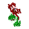
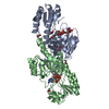
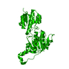
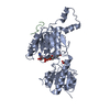
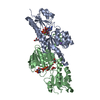
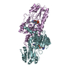
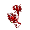
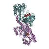
A: C-terminal-binding protein 2
B: C-terminal-binding protein 2
C: C-terminal-binding protein 2
D: C-terminal-binding protein 2
E: C-terminal-binding protein 2
F: C-terminal-binding protein 2
G: C-terminal-binding protein 2
H: C-terminal-binding protein 2
hetero molecules


Theoretical massNumber of molelcules
Total (without water)314,65124
Polymers308,1598
Non-polymers6,49316
Water3,693205
1
A: C-terminal-binding protein 2
B: C-terminal-binding protein 2
hetero molecules


Theoretical massNumber of molelcules
Total (without water)78,6636
Polymers77,0402
Non-polymers1,6234
Water362
TypeNameSymmetry operationNumber
identity operation1_555x,y,z1
Buried area8220 Å2
ΔGint-46 kcal/mol
Surface area24410 Å2
MethodPISA
2
C: C-terminal-binding protein 2
D: C-terminal-binding protein 2
hetero molecules


Theoretical massNumber of molelcules
Total (without water)78,6636
Polymers77,0402
Non-polymers1,6234
Water362
TypeNameSymmetry operationNumber
identity operation1_555x,y,z1
Buried area8320 Å2
ΔGint-44 kcal/mol
Surface area24290 Å2
MethodPISA
3
E: C-terminal-binding protein 2
F: C-terminal-binding protein 2
hetero molecules


Theoretical massNumber of molelcules
Total (without water)78,6636
Polymers77,0402
Non-polymers1,6234
Water362
TypeNameSymmetry operationNumber
identity operation1_555x,y,z1
Buried area8040 Å2
ΔGint-46 kcal/mol
Surface area24340 Å2
MethodPISA
4
G: C-terminal-binding protein 2
H: C-terminal-binding protein 2
hetero molecules


Theoretical massNumber of molelcules
Total (without water)78,6636
Polymers77,0402
Non-polymers1,6234
Water362
TypeNameSymmetry operationNumber
identity operation1_555x,y,z1
Buried area8020 Å2
ΔGint-46 kcal/mol
Surface area24300 Å2
MethodPISA
Unit cell
Length a, b, c (Å)86.151, 140.605, 135.126
Angle α, β, γ (deg.)90.000, 97.870, 90.000
Int Tables number4
Space group name H-MP1211

-
Components

#1: Protein
C-terminal-binding protein 2 / CtBP2


Mass: 38519.828 Da / Num. of mol.: 8 / Fragment: UNP residues 31-362
Source method: isolated from a genetically manipulated source
Source: (gene. exp.) Homo sapiens (human) / Gene: CTBP2 / Plasmid: pET28A / Production host: Escherichia coli (E. coli) / Strain (production host): BL21(DE3) Codon Plus RIL / References: UniProt: P56545
#2: Chemical
ChemComp-NAD / NICOTINAMIDE-ADENINE-DINUCLEOTIDE / Nicotinamide adenine dinucleotide


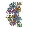
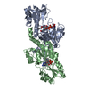
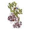
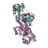
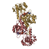
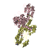
Mass: 663.425 Da / Num. of mol.: 8 / Source method: obtained synthetically / Formula: C21H27N7O14P2 / Comment: NAD*YM
#3: Chemical
ChemComp-KMT / 4-(METHYLSULFANYL)-2-OXOBUTANOIC ACID


Mass: 148.180 Da / Num. of mol.: 8 / Source method: obtained synthetically / Formula: C5H8O3S
#4: Water ChemComp-HOH / water / Water


Mass: 18.015 Da / Num. of mol.: 205 / Source method: isolated from a natural source / Formula: H2O

-
Experimental details

-
Experiment

ExperimentMethod: X-RAY DIFFRACTION / Number of used crystals: 1

-
Sample preparation

CrystalDensity Matthews: 2.63 Å3/Da / Density % sol: 53.25 %
Crystal growTemperature: 298 K / Method: vapor diffusion, hanging drop / pH: 7
Details: 200 mM potassium nitrate, 15-20% PEG 3350, 100 mM bis tris propane, pH 7.0, VAPOR DIFFUSION, HANGING DROP, temperature 298K

-
Data collection

DiffractionMean temperature: 100 K
Diffraction sourceSource: SYNCHROTRON / Site: APS / Beamline: 14-BM-C / Wavelength: 0.99 Å
DetectorType: ADSC QUANTUM 315 / Detector: CCD / Date: Jun 30, 2011
RadiationMonochromator: Bent Ge(111) monochromator / Protocol: SINGLE WAVELENGTH / Monochromatic (M) / Laue (L): M / Scattering type: x-ray
Radiation wavelengthWavelength: 0.99 Å / Relative weight: 1
ReflectionRedundancy: 4.1 % / Av σ(I) over netI: 13.24 / Number: 298092 / Rmerge(I) obs: 0.093 / Χ2: 1 / D res high: 2.86 Å / D res low: 50 Å / Num. obs: 73374 / % possible obs: 99.4
Diffraction reflection shell
Highest resolution (Å)Lowest resolution (Å)% possible obs (%)IDRmerge(I) obsChi squaredRedundancy
6.165098.610.0520.9964
4.896.1699.910.0710.9964
4.274.8999.910.0730.9944.1
3.884.2799.910.0861.0024.1
3.63.8899.910.1040.9924.1
3.393.699.710.1250.9974.1
3.223.3999.610.1671.0054.1
3.083.229910.2331.0064.1
2.963.0898.710.3130.9994.1
2.862.9698.810.3891.0034
ReflectionResolution: 2.86→50 Å / Num. all: 73837 / Num. obs: 73374 / % possible obs: 99.4 % / Observed criterion σ(I): -3 / Redundancy: 4.1 % / Rmerge(I) obs: 0.093 / Χ2: 0.999 / Net I/σ(I): 10
Reflection shell
Resolution (Å)Redundancy (%)Rmerge(I) obsNum. unique allΧ2Diffraction-ID% possible all
2.86-2.9640.38972631.003198.8
2.96-3.084.10.31372530.999198.7
3.08-3.224.10.23372761.006199
3.22-3.394.10.16773191.005199.6
3.39-3.64.10.12573550.997199.7
3.6-3.884.10.10473390.992199.9
3.88-4.274.10.08673681.002199.9
4.27-4.894.10.07373770.994199.9
4.89-6.1640.07174290.996199.9
6.16-5040.05273950.996198.6

-
Phasing

PhasingMethod: molecular replacement
Phasing MRModel details: Phaser MODE: MR_AUTO
Highest resolutionLowest resolution
Rotation2.86 Å32.3 Å
Translation2.86 Å32.3 Å

-
Processing

Software
NameVersionClassificationNB
DENZOdata reduction
SCALEPACKdata scaling
PHASER2.5.1phasing
PHENIX1.8.1_1168refinement
PDB_EXTRACT3.11data extraction
ADSCQuantumdata collection
HKL-2000data reduction
RefinementMethod to determine structure: MOLECULAR REPLACEMENT / Resolution: 2.86→32.303 Å / Occupancy max: 1 / Occupancy min: 0.37 / SU ML: 0.36 / σ(F): 1.34 / Phase error: 28.7 / Stereochemistry target values: ML
RfactorNum. reflection% reflectionSelection details
Rfree0.2506 3692 5.04 %Random
Rwork0.2139 ---
obs0.2158 73303 99.2 %-
all-73327 --
Solvent computationShrinkage radii: 0.9 Å / VDW probe radii: 1.11 Å / Solvent model: FLAT BULK SOLVENT MODEL
Displacement parametersBiso max: 82.94 Å2 / Biso mean: 52.6922 Å2 / Biso min: 29.8 Å2
Refinement stepCycle: LAST / Resolution: 2.86→32.303 Å
ProteinNucleic acidLigandSolventTotal
Num. atoms19394 0 424 205 20023
Refine LS restraints
Refine-IDTypeDev idealNumber
X-RAY DIFFRACTIONf_bond_d0.00920200
X-RAY DIFFRACTIONf_angle_d1.21927553
X-RAY DIFFRACTIONf_chiral_restr0.0573243
X-RAY DIFFRACTIONf_plane_restr0.0073581
X-RAY DIFFRACTIONf_dihedral_angle_d17.3417111
LS refinement shell

Refine-ID: X-RAY DIFFRACTION / Total num. of bins used: 26

Resolution (Å)Rfactor RfreeNum. reflection RfreeRfactor RworkNum. reflection RworkNum. reflection all% reflection obs (%)
2.86-2.89410.33191210.31212545266693
2.8941-2.93370.32451280.292648277699
2.9337-2.97560.34261370.28192666280399
2.9756-3.020.3291480.28452651279999
3.02-3.06710.281520.28132655280799
3.0671-3.11740.29591530.26482608276199
3.1174-3.17110.38141240.26782681280599
3.1711-3.22870.28981170.26982681279899
3.2287-3.29080.35031490.26792673282299
3.2908-3.35790.28941340.243126712805100
3.3579-3.43080.31841430.242227232866100
3.4308-3.51050.2791510.232126352786100
3.5105-3.59820.28531460.236327082854100
3.5982-3.69530.24021460.220626662812100
3.6953-3.80390.30041260.215327092835100
3.8039-3.92650.25381600.213826892849100
3.9265-4.06660.21051630.196626552818100
4.0666-4.22910.22831450.19526992844100
4.2291-4.42110.23581330.187126802813100
4.4211-4.65350.20731570.175326892846100
4.6535-4.94410.21181290.176126982827100
4.9441-5.32430.22191640.179126882852100
5.3243-5.85720.23941380.203427322870100
5.8572-6.69820.22961460.217127222868100
6.6982-8.41430.20611360.181727262862100
8.4143-32.30460.20581460.18462713285998

+
About Yorodumi

-
News

-
Feb 9, 2022. New format data for meta-information of EMDB entries

New format data for meta-information of EMDB entries

  • Version 3 of the EMDB header file is now the official format.
  • The previous official version 1.9 will be removed from the archive.

Related info.:EMDB header

External links:wwPDB to switch to version 3 of the EMDB data model

-
Aug 12, 2020. Covid-19 info

Covid-19 info

URL: https://pdbj.org/emnavi/covid19.php

New page: Covid-19 featured information page in EM Navigator.

Related info.:Covid-19 info / Mar 5, 2020. Novel coronavirus structure data

+
Mar 5, 2020. Novel coronavirus structure data

Novel coronavirus structure data

Related info.:Yorodumi Speices / Aug 12, 2020. Covid-19 info

External links:COVID-19 featured content - PDBj / Molecule of the Month (242):Coronavirus Proteases

+
Jan 31, 2019. EMDB accession codes are about to change! (news from PDBe EMDB page)

EMDB accession codes are about to change! (news from PDBe EMDB page)

  • The allocation of 4 digits for EMDB accession codes will soon come to an end. Whilst these codes will remain in use, new EMDB accession codes will include an additional digit and will expand incrementally as the available range of codes is exhausted. The current 4-digit format prefixed with “EMD-” (i.e. EMD-XXXX) will advance to a 5-digit format (i.e. EMD-XXXXX), and so on. It is currently estimated that the 4-digit codes will be depleted around Spring 2019, at which point the 5-digit format will come into force.
  • The EM Navigator/Yorodumi systems omit the EMD- prefix.

Related info.:Q: What is EMD? / ID/Accession-code notation in Yorodumi/EM Navigator

External links:EMDB Accession Codes are Changing Soon! / Contact to PDBj

+
Jul 12, 2017. Major update of PDB

Major update of PDB

  • wwPDB released updated PDB data conforming to the new PDBx/mmCIF dictionary.
  • This is a major update changing the version number from 4 to 5, and with Remediation, in which all the entries are updated.
  • In this update, many items about electron microscopy experimental information are reorganized (e.g. em_software).
  • Now, EM Navigator and Yorodumi are based on the updated data.

External links:wwPDB Remediation / Enriched Model Files Conforming to OneDep Data Standards Now Available in the PDB FTP Archive

-
Yorodumi

Thousand views of thousand structures

  • Yorodumi is a browser for structure data from EMDB, PDB, SASBDB, etc.
  • This page is also the successor to EM Navigator detail page, and also detail information page/front-end page for Omokage search.
  • The word "yorodu" (or yorozu) is an old Japanese word meaning "ten thousand". "mi" (miru) is to see.

Related info.:EMDB / PDB / SASBDB / Comparison of 3 databanks / Yorodumi Search / Aug 31, 2016. New EM Navigator & Yorodumi / Yorodumi Papers / Jmol/JSmol / Function and homology information / Changes in new EM Navigator and Yorodumi

Read more