 Movie
Movie Controller


+ Open data

- Basic information
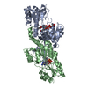
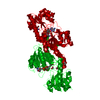
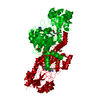
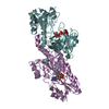
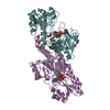
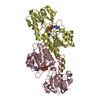
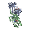
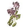
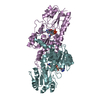
| Entry | Database: PDB / ID: 6v8a | ||||||
|---|---|---|---|---|---|---|---|
| Title | Human CtBP1 (28-375) in complex with AMP | ||||||
 Components Components | C-terminal-binding protein 1 | ||||||
 Keywords Keywords | TRANSCRIPTION / co-transcriptional factor | ||||||
| Function / homology |  Function and homology information Function and homology informationSignaling by TCF7L2 mutants / Repression of WNT target genes / synaptic vesicle clustering / presynaptic active zone cytoplasmic component / Oxidoreductases; Acting on the CH-OH group of donors; With NAD+ or NADP+ as acceptor / oxidoreductase activity, acting on the CH-OH group of donors, NAD or NADP as acceptor / lncRNA binding / white fat cell differentiation / synaptic vesicle endocytosis / Notch signaling pathway ...Signaling by TCF7L2 mutants / Repression of WNT target genes / synaptic vesicle clustering / presynaptic active zone cytoplasmic component / Oxidoreductases; Acting on the CH-OH group of donors; With NAD+ or NADP+ as acceptor / oxidoreductase activity, acting on the CH-OH group of donors, NAD or NADP as acceptor / lncRNA binding / white fat cell differentiation / synaptic vesicle endocytosis / Notch signaling pathway / transcription repressor complex / SUMOylation of transcription cofactors / viral genome replication / transcription corepressor binding / transcription coregulator binding / Deactivation of the beta-catenin transactivating complex / Negative Regulation of CDH1 Gene Transcription / GABA-ergic synapse / NAD binding / transcription corepressor activity / DNA-binding transcription factor binding / RNA polymerase II-specific DNA-binding transcription factor binding / protein phosphorylation / transcription coactivator activity / regulation of cell cycle / negative regulation of cell population proliferation / protein domain specific binding / negative regulation of DNA-templated transcription / chromatin binding / regulation of transcription by RNA polymerase II / glutamatergic synapse / negative regulation of transcription by RNA polymerase II / nucleoplasm / identical protein binding / nucleus Similarity search - Function | ||||||
| Biological species |  Homo sapiens (human) Homo sapiens (human) | ||||||
| Method |  X-RAY DIFFRACTION / MOLECULAR REPLACEMENT / Resolution: 2.35 Å X-RAY DIFFRACTION / MOLECULAR REPLACEMENT / Resolution: 2.35 Å | ||||||
 Authors Authors | Royer, W.E. | ||||||
| Funding support |  United States, 1items United States, 1items
| ||||||
 Citation Citation | Journal: J.Biol.Chem. / Year: 2021 Title: NAD(H) phosphates mediate tetramer assembly of human C-terminal binding protein (CtBP). Authors: Nichols, J.C. / Schiffer, C.A. / Royer Jr., W.E. | ||||||
| History |
|
- Structure visualization
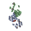
| Structure viewer | Molecule: MolmilJmol/JSmol |
|---|
- Downloads & links
-Download
| PDBx/mmCIF format | 6v8a.cif.gz | 183.4 KB | Display | PDBx/mmCIF format |
|---|---|---|---|---|
| PDB format | pdb6v8a.ent.gz | 118.4 KB | Display | PDB format |
| PDBx/mmJSON format | 6v8a.json.gz | Tree view | PDBx/mmJSON format | |
| Others |  Other downloads Other downloads |
-Validation report
| Arichive directory | https://data.pdbj.org/pub/pdb/validation_reports/v8/6v8aftp://data.pdbj.org/pub/pdb/validation_reports/v8/6v8a | HTTPS FTP |
|---|
-Related structure data
| Related structure data | 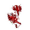 6v89C 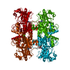 7kwmC 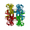 6cdfS S: Starting model for refinement C: citing same article ( |
|---|---|
| Similar structure data |
-Links
 PDBj
PDBj


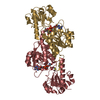
- Assembly
| Deposited unit | 
| ||||||||||||
|---|---|---|---|---|---|---|---|---|---|---|---|---|---|
| 1 | 
| ||||||||||||
| Unit cell |
| ||||||||||||
| Components on special symmetry positions |
|
-Components
| #1: Protein | Mass: 40539.066 Da / Num. of mol.: 1 / Fragment: UNP residues 28-375 / Mutation: V185T Source method: isolated from a genetically manipulated source Source: (gene. exp.) Homo sapiens (human) / Gene: CTBP1, CTBP / Production host:  References: UniProt: Q13363, Oxidoreductases; Acting on the CH-OH group of donors; With NAD+ or NADP+ as acceptor | ||||
|---|---|---|---|---|---|
| #2: Chemical | ChemComp-CA /   Mass: 40.078 Da / Num. of mol.: 1 / Source method: obtained synthetically / Formula: Ca Mass: 40.078 Da / Num. of mol.: 1 / Source method: obtained synthetically / Formula: Ca | ||||
| #3: Chemical | ChemComp-AMP /   Mass: 347.221 Da / Num. of mol.: 1 / Source method: obtained synthetically / Formula: C10H14N5O7P / Feature type: SUBJECT OF INVESTIGATION / Comment: AMP*YM Mass: 347.221 Da / Num. of mol.: 1 / Source method: obtained synthetically / Formula: C10H14N5O7P / Feature type: SUBJECT OF INVESTIGATION / Comment: AMP*YM | ||||
| #4: Chemical |   Mass: 35.453 Da / Num. of mol.: 3 / Source method: obtained synthetically / Formula: Cl Mass: 35.453 Da / Num. of mol.: 3 / Source method: obtained synthetically / Formula: Cl#5: Water | ChemComp-HOH / |  Mass: 18.015 Da / Num. of mol.: 220 / Source method: isolated from a natural source / Formula: H2O Mass: 18.015 Da / Num. of mol.: 220 / Source method: isolated from a natural source / Formula: H2OHas ligand of interest | Y | |
-Experimental details
-Experiment
| Experiment | Method: X-RAY DIFFRACTION / Number of used crystals: 1 |
|---|
- Sample preparation
| Crystal | Density Matthews: 2.32 Å3/Da / Density % sol: 47.05 % |
|---|---|
| Crystal grow | Temperature: 298 K / Method: vapor diffusion, hanging drop / pH: 7.5 Details: 100 mM HEPES, pH 7.5, 80-200 mM calcium chloride, 2-6% PEG400 |
-Data collection
| Diffraction | Mean temperature: 100 K / Serial crystal experiment: N | |||||||||||||||||||||||||||||||||||||||||||||||||||||||||||||||||||||||||||||||||||||||||||||||||||||||||||||||||||||||||||||||||||||||||||||||||||
|---|---|---|---|---|---|---|---|---|---|---|---|---|---|---|---|---|---|---|---|---|---|---|---|---|---|---|---|---|---|---|---|---|---|---|---|---|---|---|---|---|---|---|---|---|---|---|---|---|---|---|---|---|---|---|---|---|---|---|---|---|---|---|---|---|---|---|---|---|---|---|---|---|---|---|---|---|---|---|---|---|---|---|---|---|---|---|---|---|---|---|---|---|---|---|---|---|---|---|---|---|---|---|---|---|---|---|---|---|---|---|---|---|---|---|---|---|---|---|---|---|---|---|---|---|---|---|---|---|---|---|---|---|---|---|---|---|---|---|---|---|---|---|---|---|---|---|---|---|
| Diffraction source | Source: ROTATING ANODE / Type: Cu FINE FOCUS / Wavelength: 1.54178 Å | |||||||||||||||||||||||||||||||||||||||||||||||||||||||||||||||||||||||||||||||||||||||||||||||||||||||||||||||||||||||||||||||||||||||||||||||||||
| Detector | Type: RIGAKU SATURN 944 / Detector: CCD / Date: Jul 3, 2019 | |||||||||||||||||||||||||||||||||||||||||||||||||||||||||||||||||||||||||||||||||||||||||||||||||||||||||||||||||||||||||||||||||||||||||||||||||||
| Radiation | Protocol: SINGLE WAVELENGTH / Monochromatic (M) / Laue (L): M / Scattering type: x-ray | |||||||||||||||||||||||||||||||||||||||||||||||||||||||||||||||||||||||||||||||||||||||||||||||||||||||||||||||||||||||||||||||||||||||||||||||||||
| Radiation wavelength | Wavelength: 1.54178 Å / Relative weight: 1 | |||||||||||||||||||||||||||||||||||||||||||||||||||||||||||||||||||||||||||||||||||||||||||||||||||||||||||||||||||||||||||||||||||||||||||||||||||
| Reflection | Resolution: 2.35→50 Å / Num. obs: 16758 / % possible obs: 99.7 % / Redundancy: 9.4 % / Rmerge(I) obs: 0.107 / Χ2: 0.939 / Net I/σ(I): 6.4 / Num. measured all: 157772 | |||||||||||||||||||||||||||||||||||||||||||||||||||||||||||||||||||||||||||||||||||||||||||||||||||||||||||||||||||||||||||||||||||||||||||||||||||
| Reflection shell |
|
- Processing
| Software |
| |||||||||||||||||||||||||||||||||||||||||||||||||
|---|---|---|---|---|---|---|---|---|---|---|---|---|---|---|---|---|---|---|---|---|---|---|---|---|---|---|---|---|---|---|---|---|---|---|---|---|---|---|---|---|---|---|---|---|---|---|---|---|---|---|
| Refinement | Method to determine structure: MOLECULAR REPLACEMENT Starting model: PDb entry 6CDF Resolution: 2.35→31.55 Å / SU ML: 0.2759 / Cross valid method: FREE R-VALUE / σ(F): 1.34 / Phase error: 23.4615
| |||||||||||||||||||||||||||||||||||||||||||||||||
| Solvent computation | Shrinkage radii: 0.9 Å / VDW probe radii: 1.11 Å | |||||||||||||||||||||||||||||||||||||||||||||||||
| Displacement parameters | Biso mean: 40.84 Å2 | |||||||||||||||||||||||||||||||||||||||||||||||||
| Refinement step | Cycle: LAST / Resolution: 2.35→31.55 Å
| |||||||||||||||||||||||||||||||||||||||||||||||||
| Refine LS restraints |
| |||||||||||||||||||||||||||||||||||||||||||||||||
| LS refinement shell |
| |||||||||||||||||||||||||||||||||||||||||||||||||
| Refinement TLS params. | Method: refined / Origin x: 2.74466670686 Å / Origin y: 32.5396989643 Å / Origin z: -26.5986527754 Å
| |||||||||||||||||||||||||||||||||||||||||||||||||
| Refinement TLS group | Selection details: all |
