 Movie
Movie Controller

[English] 日本語
 Yorodumi
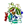
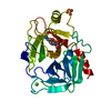
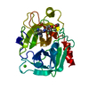
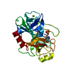
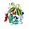
- PDB-1s85: PORCINE TRYPSIN COMPLEXED WITH P-HYDROXYMETHYL BENZAMIDINE AND BORATE -
+ Open data

- Basic information
| Entry | Database: PDB / ID: 1s85 | ||||||
|---|---|---|---|---|---|---|---|
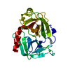
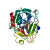
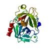
| Title | PORCINE TRYPSIN COMPLEXED WITH P-HYDROXYMETHYL BENZAMIDINE AND BORATE | ||||||
 Components Components | TRYPSIN | ||||||
 Keywords Keywords | HYDROLASE / SERINE PROTEASE | ||||||
| Function / homology |  Function and homology information Function and homology informationtrypsin / digestion / serine-type endopeptidase activity / proteolysis / : / metal ion binding Similarity search - Function | ||||||
| Biological species |  | ||||||
| Method |  X-RAY DIFFRACTION / MOLECULAR REPLACEMENT / Resolution: 2.2 Å X-RAY DIFFRACTION / MOLECULAR REPLACEMENT / Resolution: 2.2 Å | ||||||
 Authors Authors | Transue, T.R. / Krahn, J.M. / Gabel, S.A. / Derose, E.F. / London, R.E. | ||||||
 Citation Citation | Journal: Biochemistry / Year: 2004 Title: X-ray and NMR characterization of covalent complexes of trypsin, borate, and alcohols. Authors: Transue, T.R. / Krahn, J.M. / Gabel, S.A. / DeRose, E.F. / London, R.E. | ||||||
| History |
|
- Structure visualization
| Structure viewer | Molecule: MolmilJmol/JSmol |
|---|
- Downloads & links
-Download
| PDBx/mmCIF format | 1s85.cif.gz | 106 KB | Display | PDBx/mmCIF format |
|---|---|---|---|---|
| PDB format | pdb1s85.ent.gz | 85.5 KB | Display | PDB format |
| PDBx/mmJSON format | 1s85.json.gz | Tree view | PDBx/mmJSON format | |
| Others |  Other downloads Other downloads |
-Validation report
| Arichive directory | https://data.pdbj.org/pub/pdb/validation_reports/s8/1s85ftp://data.pdbj.org/pub/pdb/validation_reports/s8/1s85 | HTTPS FTP |
|---|
-Related structure data
| Related structure data | 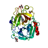 1s5sC 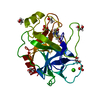 1s6fC  1s6hC 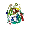 1s81C 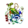 1s82C 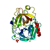 1s83C 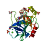 1s84C 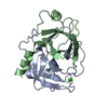 1aksS C: citing same article ( S: Starting model for refinement |
|---|---|
| Similar structure data |


-Links
 PDBj
PDBj

- Assembly
| Deposited unit | 
| ||||||||
|---|---|---|---|---|---|---|---|---|---|
| 1 |
| ||||||||
| Unit cell |
|
-Components
| #1: Protein | Mass: 23493.496 Da / Num. of mol.: 1 / Source method: isolated from a natural source / Source: (natural) |
|---|---|
| #2: Chemical | ChemComp-CA /   Mass: 40.078 Da / Num. of mol.: 1 / Source method: obtained synthetically / Formula: Ca Mass: 40.078 Da / Num. of mol.: 1 / Source method: obtained synthetically / Formula: Ca |
| #3: Chemical | ChemComp-SO4 /   Mass: 96.063 Da / Num. of mol.: 1 / Source method: obtained synthetically / Formula: SO4 Mass: 96.063 Da / Num. of mol.: 1 / Source method: obtained synthetically / Formula: SO4 |
| #4: Chemical | ChemComp-SBZ / [  Mass: 221.041 Da / Num. of mol.: 1 / Source method: obtained synthetically / Formula: C10H14BN2O3 Mass: 221.041 Da / Num. of mol.: 1 / Source method: obtained synthetically / Formula: C10H14BN2O3 |
| #5: Water | ChemComp-HOH /  Mass: 18.015 Da / Num. of mol.: 218 / Source method: isolated from a natural source / Formula: H2O Mass: 18.015 Da / Num. of mol.: 218 / Source method: isolated from a natural source / Formula: H2O |
| Has protein modification | Y |
-Experimental details
-Experiment
| Experiment | Method: X-RAY DIFFRACTION / Number of used crystals: 1 |
|---|
- Sample preparation
| Crystal | Density Matthews: 2.1 Å3/Da / Density % sol: 41.37 % | |||||||||||||||||||||||||||||||||||||||||||||||||
|---|---|---|---|---|---|---|---|---|---|---|---|---|---|---|---|---|---|---|---|---|---|---|---|---|---|---|---|---|---|---|---|---|---|---|---|---|---|---|---|---|---|---|---|---|---|---|---|---|---|---|
| Crystal grow | Temperature: 298 K / Method: vapor diffusion, sitting drop / pH: 8 Details: 1.8M MgCl2, 50mM HEPES, 5mM CaCl2, VAPOR DIFFUSION, SITTING DROP, temperature 298K | |||||||||||||||||||||||||||||||||||||||||||||||||
| Crystal grow | *PLUS Temperature: 4 ℃ / pH: 8 / Method: vapor diffusion, sitting drop | |||||||||||||||||||||||||||||||||||||||||||||||||
| Components of the solutions | *PLUS
|
-Data collection
| Diffraction | Mean temperature: 100 K |
|---|---|
| Diffraction source | Source: ROTATING ANODE / Type: RIGAKU / Wavelength: 1.5418 / Wavelength: 1.5418 Å |
| Detector | Type: RIGAKU RAXIS IV / Detector: IMAGE PLATE / Date: Jun 20, 2002 / Details: MSC/YALE DOUBLE FOCUSING MIRRORS |
| Radiation | Monochromator: MSC/YALE DOUBLE FOCUSING MIRRORS / Protocol: SINGLE WAVELENGTH / Monochromatic (M) / Laue (L): M / Scattering type: x-ray |
| Radiation wavelength | Wavelength: 1.5418 Å / Relative weight: 1 |
| Reflection | Resolution: 2.2→100 Å / Num. all: 10156 / Num. obs: 10156 / % possible obs: 96.5 % / Observed criterion σ(F): 0 / Observed criterion σ(I): -3 / Redundancy: 8.325 % / Rsym value: 0.111 / Net I/σ(I): 20.4867 |
| Reflection shell | Resolution: 2.2→2.28 Å / Redundancy: 8.72 % / Mean I/σ(I) obs: 6.391 / Num. unique all: 1038 / Rsym value: 0.377 / % possible all: 100 |
| Reflection | *PLUS Highest resolution: 2.2 Å / % possible obs: 99.8 % / Redundancy: 8.3 % / Rmerge(I) obs: 0.111 |
| Reflection shell | *PLUS Rmerge(I) obs: 0.377 / Mean I/σ(I) obs: 6.4 |
- Processing
| Software |
| ||||||||||||||||||||
|---|---|---|---|---|---|---|---|---|---|---|---|---|---|---|---|---|---|---|---|---|---|
| Refinement | Method to determine structure: MOLECULAR REPLACEMENT Starting model: PDB ENTRY 1AKS Resolution: 2.2→100 Å / Isotropic thermal model: ISOTROPIC / Cross valid method: THROUGHOUT / σ(F): 0 / Stereochemistry target values: AMBER98
| ||||||||||||||||||||
| Displacement parameters | Biso mean: 25.93 Å2
| ||||||||||||||||||||
| Refinement step | Cycle: LAST / Resolution: 2.2→100 Å
| ||||||||||||||||||||
| Refine LS restraints |
| ||||||||||||||||||||
| Refinement | *PLUS Highest resolution: 2.2 Å / % reflection Rfree: 5 % | ||||||||||||||||||||
| Solvent computation | *PLUS | ||||||||||||||||||||
| Displacement parameters | *PLUS |
