 Movie
Movie Controller

[English] 日本語
 Yorodumi
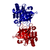
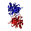
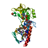
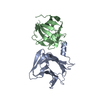
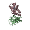
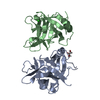
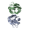
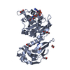
- PDB-1gx9: BOVINE BETA-LACTOGLOBULIN COMPLEXED WITH RETINOIC ACID, TRIGONAL ... -
+ Open data

- Basic information
| Entry | Database: PDB / ID: 1gx9 | ||||||
|---|---|---|---|---|---|---|---|
| Title | BOVINE BETA-LACTOGLOBULIN COMPLEXED WITH RETINOIC ACID, TRIGONAL LATTICE Z | ||||||
 Components Components | BETA-LACTOGLOBULIN | ||||||
 Keywords Keywords | LIPOCALIN / MILK / WHEY TRANSPORT / BOVINE / RETINOL-BINDING ALLERGEN / RETINOIC ACID-BINDING | ||||||
| Function / homology |  Function and homology information Function and homology informationretinol binding / long-chain fatty acid binding / extracellular region / identical protein binding Similarity search - Function | ||||||
| Biological species |  | ||||||
| Method |  X-RAY DIFFRACTION / MOLECULAR REPLACEMENT / Resolution: 2.34 Å X-RAY DIFFRACTION / MOLECULAR REPLACEMENT / Resolution: 2.34 Å | ||||||
 Authors Authors | Kontopidis, G. / Sawyer, L. | ||||||
 Citation Citation | Journal: J.Mol.Biol. / Year: 2002 Title: The Ligand-Binding Site of Bovine Beta-Lactoglobulin: Evidence for a Function? Authors: Kontopidis, G. / Holt, C. / Sawyer, L. | ||||||
| History |
|
- Structure visualization
| Structure viewer | Molecule: MolmilJmol/JSmol |
|---|
- Downloads & links
-Download
| PDBx/mmCIF format | 1gx9.cif.gz | 49.2 KB | Display | PDBx/mmCIF format |
|---|---|---|---|---|
| PDB format | pdb1gx9.ent.gz | 34.5 KB | Display | PDB format |
| PDBx/mmJSON format | 1gx9.json.gz | Tree view | PDBx/mmJSON format | |
| Others |  Other downloads Other downloads |
-Validation report
| Summary document | 1gx9_validation.pdf.gz | 428.5 KB | Display | wwPDB validaton report |
|---|---|---|---|---|
| Full document | 1gx9_full_validation.pdf.gz | 437.3 KB | Display | |
| Data in XML | 1gx9_validation.xml.gz | 6.1 KB | Display | |
| Data in CIF | 1gx9_validation.cif.gz | 9 KB | Display | |
| Arichive directory | https://data.pdbj.org/pub/pdb/validation_reports/gx/1gx9ftp://data.pdbj.org/pub/pdb/validation_reports/gx/1gx9 | HTTPS FTP |
-Related structure data
| Related structure data |  1gx8C  1gxaC  1b0oS S: Starting model for refinement C: citing same article ( |
|---|---|
| Similar structure data |


-Links
 PDBj
PDBj

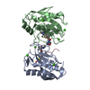
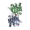
- Assembly
| Deposited unit | 
| ||||||||
|---|---|---|---|---|---|---|---|---|---|
| 1 | 
| ||||||||
| Unit cell |
| ||||||||
| Components on special symmetry positions |
| ||||||||
| Details | THE DIMER GIVEN HERE IS PHYSIOLOGICAL DIMERTHE STRAND AA10 CREATES A CONTINUOUS BETA SHEET BETWEENRESIDUES 147-150 IN THE DIMER |
-Components
| #1: Protein | Mass: 18301.174 Da / Num. of mol.: 1 / Source method: isolated from a natural source Details: PROTEIN PURCHASED FROM SIGMA CHEMICALS, CAT.NO. L8005 Source: (natural) |
|---|---|
| #2: Chemical | ChemComp-REA / 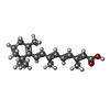  Mass: 300.435 Da / Num. of mol.: 1 / Source method: obtained synthetically / Formula: C20H28O2 Mass: 300.435 Da / Num. of mol.: 1 / Source method: obtained synthetically / Formula: C20H28O2 |
| #3: Water | ChemComp-HOH /  Mass: 18.015 Da / Num. of mol.: 139 / Source method: isolated from a natural source / Formula: H2O Mass: 18.015 Da / Num. of mol.: 139 / Source method: isolated from a natural source / Formula: H2O |
| Has protein modification | Y |
-Experimental details
-Experiment
| Experiment | Method: X-RAY DIFFRACTION / Number of used crystals: 1 |
|---|
- Sample preparation
| Crystal | Density Matthews: 2.495 Å3/Da / Density % sol: 51.3 % | ||||||||||||||||||||||||||||||
|---|---|---|---|---|---|---|---|---|---|---|---|---|---|---|---|---|---|---|---|---|---|---|---|---|---|---|---|---|---|---|---|
| Crystal grow | Temperature: 290 K / pH: 7.3 Details: 17 DEG C, 8MUL BLG-RET COMPLEX 20MM TRIS, PH 8 + 8MUL NA CITRATE 1.25M, 0.1M HEPES, PH7.3, PER DROP | ||||||||||||||||||||||||||||||
| Crystal grow | *PLUS Temperature: 17 ℃ / pH: 8 / Method: vapor diffusion, hanging drop | ||||||||||||||||||||||||||||||
| Components of the solutions | *PLUS
|
-Data collection
| Diffraction | Mean temperature: 100 K |
|---|---|
| Diffraction source | Source: ROTATING ANODE / Type: ENRAF-NONIUS FR571 / Wavelength: 1.542 |
| Detector | Type: RAYONIX MX340-HS / Detector: CCD / Details: COLLIMATOR |
| Radiation | Monochromator: GRAPHITE / Protocol: SINGLE WAVELENGTH / Monochromatic (M) / Laue (L): M / Scattering type: x-ray |
| Radiation wavelength | Wavelength: 1.542 Å / Relative weight: 1 |
| Reflection | Resolution: 2.34→15 Å / Num. obs: 8213 / % possible obs: 99.6 % / Redundancy: 16.8 % / Rmerge(I) obs: 0.057 / Net I/σ(I): 23.3 |
| Reflection shell | Rmerge(I) obs: 0.25 / % possible all: 100 |
| Reflection | *PLUS Lowest resolution: 15 Å / Num. measured all: 138409 |
| Reflection shell | *PLUS % possible obs: 100 % / Rmerge(I) obs: 0.25 |
- Processing
| Software |
| |||||||||||||||||||||||||||||||||
|---|---|---|---|---|---|---|---|---|---|---|---|---|---|---|---|---|---|---|---|---|---|---|---|---|---|---|---|---|---|---|---|---|---|---|
| Refinement | Method to determine structure: MOLECULAR REPLACEMENT Starting model: 1B0O Resolution: 2.34→15 Å / Num. parameters: 5758 / Num. restraintsaints: 5312 / Cross valid method: FREE R-VALUE / σ(F): 0 / Stereochemistry target values: ENGH AND HUBER / Details: THE RESIDUE, LEU 1 IS NOT OBSERVED
| |||||||||||||||||||||||||||||||||
| Solvent computation | Solvent model: MOEWS & KRETSINGER, J.MOL.BIOL.91(1973)201-2 | |||||||||||||||||||||||||||||||||
| Refine analyze | Num. disordered residues: 5 / Occupancy sum hydrogen: 0 / Occupancy sum non hydrogen: 1439.5 | |||||||||||||||||||||||||||||||||
| Refinement step | Cycle: LAST / Resolution: 2.34→15 Å
| |||||||||||||||||||||||||||||||||
| Refine LS restraints |
| |||||||||||||||||||||||||||||||||
| Software | *PLUS Name: SHELXL / Version: 97 / Classification: refinement | |||||||||||||||||||||||||||||||||
| Refinement | *PLUS % reflection Rfree: 5 % / Rfactor all: 0.227 / Rfactor obs: 0.298 / Rfactor Rfree: 0.227 | |||||||||||||||||||||||||||||||||
| Solvent computation | *PLUS | |||||||||||||||||||||||||||||||||
| Displacement parameters | *PLUS | |||||||||||||||||||||||||||||||||
| Refine LS restraints | *PLUS Type: s_bond_d / Dev ideal: 0.005 |
