 Movie
Movie Controller

[English] 日本語
 Yorodumi
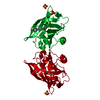
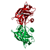
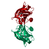
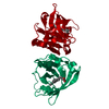
- PDB-1bso: 12-BROMODODECANOIC ACID BINDS INSIDE THE CALYX OF BOVINE BETA-LAC... -
+ Open data

- Basic information
| Entry | Database: PDB / ID: 1bso | ||||||
|---|---|---|---|---|---|---|---|
| Title | 12-BROMODODECANOIC ACID BINDS INSIDE THE CALYX OF BOVINE BETA-LACTOGLOBULIN | ||||||
 Components Components | PROTEIN (BOVINE BETA-LACTOGLOBULIN A) | ||||||
 Keywords Keywords | TRANSPORT PROTEIN / BETA-LACTOGLOBULIN / LIGAND BINDING / X-RAY CRYSTAL STRUCTURE | ||||||
| Function / homology |  Function and homology information Function and homology informationretinol binding / long-chain fatty acid binding / extracellular region / identical protein binding Similarity search - Function | ||||||
| Biological species |  | ||||||
| Method |  X-RAY DIFFRACTION / MOLECULAR REPLACEMENT / Resolution: 2.23 Å X-RAY DIFFRACTION / MOLECULAR REPLACEMENT / Resolution: 2.23 Å | ||||||
 Authors Authors | Qin, B.Y. / Creamer, L.K. / Baker, E.N. / Jameson, G.B. | ||||||
 Citation Citation | Journal: FEBS Lett. / Year: 1998 Title: 12-Bromododecanoic acid binds inside the calyx of bovine beta-lactoglobulin. Authors: Qin, B.Y. / Creamer, L.K. / Baker, E.N. / Jameson, G.B. | ||||||
| History |
|
- Structure visualization
| Structure viewer | Molecule: MolmilJmol/JSmol |
|---|
- Downloads & links
-Download
| PDBx/mmCIF format | 1bso.cif.gz | 46.9 KB | Display | PDBx/mmCIF format |
|---|---|---|---|---|
| PDB format | pdb1bso.ent.gz | 32.7 KB | Display | PDB format |
| PDBx/mmJSON format | 1bso.json.gz | Tree view | PDBx/mmJSON format | |
| Others |  Other downloads Other downloads |
-Validation report
| Arichive directory | https://data.pdbj.org/pub/pdb/validation_reports/bs/1bsoftp://data.pdbj.org/pub/pdb/validation_reports/bs/1bso | HTTPS FTP |
|---|
-Related structure data
| Similar structure data |
|---|


-Links
 PDBj
PDBj

- Assembly
| Deposited unit | 
| ||||||||
|---|---|---|---|---|---|---|---|---|---|
| 1 | 
| ||||||||
| Unit cell |
|
-Components
| #1: Protein | Mass: 18387.264 Da / Num. of mol.: 1 / Source method: isolated from a natural source / Source: (natural) |
|---|---|
| #2: Chemical | ChemComp-BRC / 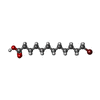  Mass: 279.214 Da / Num. of mol.: 1 / Source method: obtained synthetically / Formula: C12H23BrO2 Mass: 279.214 Da / Num. of mol.: 1 / Source method: obtained synthetically / Formula: C12H23BrO2 |
| #3: Water | ChemComp-HOH /  Mass: 18.015 Da / Num. of mol.: 66 / Source method: isolated from a natural source / Formula: H2O Mass: 18.015 Da / Num. of mol.: 66 / Source method: isolated from a natural source / Formula: H2O |
| Has protein modification | Y |
-Experimental details
-Experiment
| Experiment | Method: X-RAY DIFFRACTION / Number of used crystals: 1 |
|---|
- Sample preparation
| Crystal | Density Matthews: 2.62 Å3/Da / Density % sol: 35 % / Description: DATA WERE COLLECTED USING OSCILLATION METHOD | ||||||||||||||||||||||||||||||||||||||||||
|---|---|---|---|---|---|---|---|---|---|---|---|---|---|---|---|---|---|---|---|---|---|---|---|---|---|---|---|---|---|---|---|---|---|---|---|---|---|---|---|---|---|---|---|
| Crystal grow | pH: 7.3 Details: PROTEIN WAS CRYSTALLIZED FROM AMMONIUM SULFATE, pH 7.3 | ||||||||||||||||||||||||||||||||||||||||||
| Crystal grow | *PLUS Method: vapor diffusion, hanging drop | ||||||||||||||||||||||||||||||||||||||||||
| Components of the solutions | *PLUS
|
-Data collection
| Diffraction | Mean temperature: 298 K |
|---|---|
| Diffraction source | Source: ROTATING ANODE / Type: RIGAKU RU200 / Wavelength: 1.5418 |
| Detector | Type: RIGAKU / Detector: IMAGE PLATE / Date: Mar 15, 1998 / Details: MIRRORS |
| Radiation | Monochromator: NI / Protocol: SINGLE WAVELENGTH / Monochromatic (M) / Laue (L): M / Scattering type: x-ray |
| Radiation wavelength | Wavelength: 1.5418 Å / Relative weight: 1 |
| Reflection | Resolution: 2.23→15 Å / Num. obs: 9695 / % possible obs: 99.5 % / Observed criterion σ(I): 2 / Redundancy: 2.62 % / Biso Wilson estimate: 45.6 Å2 / Rmerge(I) obs: 0.048 / Rsym value: 0.048 / Net I/σ(I): 18.73 |
| Reflection shell | Resolution: 2.23→2.28 Å / Redundancy: 2.64 % / Rmerge(I) obs: 0.548 / Mean I/σ(I) obs: 2.04 / Rsym value: 0.548 / % possible all: 98.75 |
| Reflection | *PLUS Lowest resolution: 15 Å |
| Reflection shell | *PLUS % possible obs: 98.7 % / Mean I/σ(I) obs: 2 |
- Processing
| Software |
| ||||||||||||||||||||||||||||||||||||||||||||||||||||||||||||
|---|---|---|---|---|---|---|---|---|---|---|---|---|---|---|---|---|---|---|---|---|---|---|---|---|---|---|---|---|---|---|---|---|---|---|---|---|---|---|---|---|---|---|---|---|---|---|---|---|---|---|---|---|---|---|---|---|---|---|---|---|---|
| Refinement | Method to determine structure: MOLECULAR REPLACEMENT Starting model: BLGA IN LATTICE Z AT PH 7.1 Resolution: 2.23→15 Å / Rfactor Rfree error: 0.013 / Data cutoff high absF: 100000 / Data cutoff low absF: 0.01 / Isotropic thermal model: OVERALL / Cross valid method: THROUGHOUT / σ(F): 0
| ||||||||||||||||||||||||||||||||||||||||||||||||||||||||||||
| Displacement parameters | Biso mean: 43 Å2
| ||||||||||||||||||||||||||||||||||||||||||||||||||||||||||||
| Refine analyze |
| ||||||||||||||||||||||||||||||||||||||||||||||||||||||||||||
| Refinement step | Cycle: LAST / Resolution: 2.23→15 Å
| ||||||||||||||||||||||||||||||||||||||||||||||||||||||||||||
| Refine LS restraints |
| ||||||||||||||||||||||||||||||||||||||||||||||||||||||||||||
| LS refinement shell | Resolution: 2.23→2.33 Å / Rfactor Rfree error: 0.04 / Total num. of bins used: 8
| ||||||||||||||||||||||||||||||||||||||||||||||||||||||||||||
| Xplor file |
| ||||||||||||||||||||||||||||||||||||||||||||||||||||||||||||
| Refinement | *PLUS Num. reflection obs: 9184 / Num. reflection Rfree: 511 / Rfactor obs: 0.234 / Rfactor Rfree: 0.276 / Rfactor Rwork: 0.234 | ||||||||||||||||||||||||||||||||||||||||||||||||||||||||||||
| Solvent computation | *PLUS | ||||||||||||||||||||||||||||||||||||||||||||||||||||||||||||
| Displacement parameters | *PLUS | ||||||||||||||||||||||||||||||||||||||||||||||||||||||||||||
| Refine LS restraints | *PLUS
|
