 Movie
Movie Controller


+ Open data

- Basic information
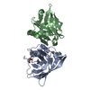
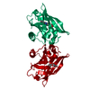
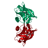
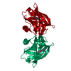
| Entry | Database: PDB / ID: 1b0o | ||||||
|---|---|---|---|---|---|---|---|
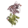
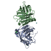
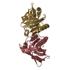
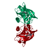
| Title | BOVINE BETA-LACTOGLOBULIN COMPLEXED WITH PALMITATE, LATTICE Z | ||||||
 Components Components | BETA-LACTOGLOBULIN | ||||||
 Keywords Keywords | LIPOCALIN / MILK WHEY PROTEIN / BOVINE / PALMITATE-BINDING | ||||||
| Function / homology |  Function and homology information Function and homology informationretinol binding / long-chain fatty acid binding / extracellular region / identical protein binding Similarity search - Function | ||||||
| Biological species |  | ||||||
| Method |  X-RAY DIFFRACTION / MOLECULAR REPLACEMENT / Resolution: 2.5 Å X-RAY DIFFRACTION / MOLECULAR REPLACEMENT / Resolution: 2.5 Å | ||||||
 Authors Authors | Wu, S.-Y. / Sawyer, L. | ||||||
 Citation Citation | Journal: J.Biol.Chem. / Year: 1999 Title: beta-lactoglobulin binds palmitate within its central cavity. Authors: Wu, S.Y. / Perez, M.D. / Puyol, P. / Sawyer, L. #1: Journal: Biochemistry / Year: 1998Title: Structural Basis of the Tanford Transition of Bovine Beta-Lactoglobulin Authors: Qin, B.Y. / Bewley, M.C. / Creamer, L.K. / Baker, H.M. / Baker, E.N. / Jameson, G.B. #2: Journal: Structure / Year: 1997Title: Bovine Beta-Lactoglobulin at 1.8 A Resolution--Still an Enigmatic Lipocalin Authors: Brownlow, S. / Morais Cabral, J.H. / Cooper, R. / Flower, D.R. / Yewdall, S.J. / Polikarpov, I. / North, A.C. / Sawyer, L. #3: Journal: FEBS Lett. / Year: 1998Title: 12-Bromododecanoic Acid Binds Inside the Calyx of Bovine Beta-Lactoglobulin Authors: Qin, B.Y. / Creamer, L.K. / Baker, E.N. / Jameson, G.B. | ||||||
| History |
|
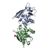
- Structure visualization
| Structure viewer | Molecule: MolmilJmol/JSmol |
|---|
- Downloads & links
-Download
| PDBx/mmCIF format | 1b0o.cif.gz | 48.2 KB | Display | PDBx/mmCIF format |
|---|---|---|---|---|
| PDB format | pdb1b0o.ent.gz | 33.6 KB | Display | PDB format |
| PDBx/mmJSON format | 1b0o.json.gz | Tree view | PDBx/mmJSON format | |
| Others |  Other downloads Other downloads |
-Validation report
| Arichive directory | https://data.pdbj.org/pub/pdb/validation_reports/b0/1b0oftp://data.pdbj.org/pub/pdb/validation_reports/b0/1b0o | HTTPS FTP |
|---|
-Related structure data
| Related structure data |  1bebS S: Starting model for refinement |
|---|---|
| Similar structure data |


-Links
 PDBj
PDBj


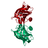
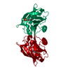
- Assembly
| Deposited unit | 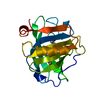
| ||||||||
|---|---|---|---|---|---|---|---|---|---|
| 1 | 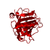
| ||||||||
| Unit cell |
|
-Components
| #1: Protein | Mass: 18301.174 Da / Num. of mol.: 1 / Source method: isolated from a natural source Details: PROTEIN PURCHASED FROM SIGMA CHEMICALS, LOT 13H7150 Source: (natural) |
|---|---|
| #2: Chemical | ChemComp-PLM /   Mass: 256.424 Da / Num. of mol.: 1 / Source method: obtained synthetically / Formula: C16H32O2 Mass: 256.424 Da / Num. of mol.: 1 / Source method: obtained synthetically / Formula: C16H32O2 |
| #3: Water | ChemComp-HOH /  Mass: 18.015 Da / Num. of mol.: 105 / Source method: isolated from a natural source / Formula: H2O Mass: 18.015 Da / Num. of mol.: 105 / Source method: isolated from a natural source / Formula: H2O |
| Has protein modification | Y |
-Experimental details
-Experiment
| Experiment | Method: X-RAY DIFFRACTION / Number of used crystals: 1 |
|---|
- Sample preparation
| Crystal | Density Matthews: 2.52 Å3/Da / Density % sol: 50.78 % | ||||||||||||||||||||||||||||||||||||
|---|---|---|---|---|---|---|---|---|---|---|---|---|---|---|---|---|---|---|---|---|---|---|---|---|---|---|---|---|---|---|---|---|---|---|---|---|---|
| Crystal grow | pH: 7.5 / Details: pH 7.5 | ||||||||||||||||||||||||||||||||||||
| Crystal grow | *PLUS Method: vapor diffusion, sitting drop | ||||||||||||||||||||||||||||||||||||
| Components of the solutions | *PLUS
|
-Data collection
| Diffraction | Mean temperature: 100 K |
|---|---|
| Diffraction source | Source: ROTATING ANODE / Type: ENRAF-NONIUS FR571 / Wavelength: 1.5418 |
| Detector | Type: MARRESEARCH / Detector: IMAGE PLATE / Date: Nov 21, 1997 / Details: COLLIMATOR |
| Radiation | Monochromator: GRAPHITE(002) / Monochromatic (M) / Laue (L): M / Scattering type: x-ray |
| Radiation wavelength | Wavelength: 1.5418 Å / Relative weight: 1 |
| Reflection | Resolution: 2.5→20 Å / Num. obs: 6888 / % possible obs: 99.9 % / Redundancy: 14.1 % / Rmerge(I) obs: 0.073 / Net I/σ(I): 21.2 |
| Reflection shell | Resolution: 2.5→2.54 Å / Redundancy: 8.16 % / Rmerge(I) obs: 0.366 / Mean I/σ(I) obs: 3.95 / % possible all: 100 |
| Reflection | *PLUS Num. measured all: 97142 |
| Reflection shell | *PLUS % possible obs: 100 % |
- Processing
| Software |
| |||||||||||||||||||||||||||||||||
|---|---|---|---|---|---|---|---|---|---|---|---|---|---|---|---|---|---|---|---|---|---|---|---|---|---|---|---|---|---|---|---|---|---|---|
| Refinement | Method to determine structure: MOLECULAR REPLACEMENT Starting model: 1BEB Resolution: 2.5→10 Å / Num. parameters: 5583 / Num. restraintsaints: 5396 / Cross valid method: FREE R / σ(F): 0 / Stereochemistry target values: ENGH AND HUBER
| |||||||||||||||||||||||||||||||||
| Refine analyze | Num. disordered residues: 0 / Occupancy sum hydrogen: 0 / Occupancy sum non hydrogen: 1395 | |||||||||||||||||||||||||||||||||
| Refinement step | Cycle: LAST / Resolution: 2.5→10 Å
| |||||||||||||||||||||||||||||||||
| Refine LS restraints |
| |||||||||||||||||||||||||||||||||
| Software | *PLUS Name: SHELXL-97 / Classification: refinement | |||||||||||||||||||||||||||||||||
| Refinement | *PLUS | |||||||||||||||||||||||||||||||||
| Solvent computation | *PLUS | |||||||||||||||||||||||||||||||||
| Displacement parameters | *PLUS Biso mean: 47.17 Å2 |
