 ムービー
ムービー コントローラー


+ データを開く

- 基本情報
基本情報
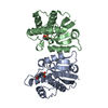
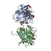
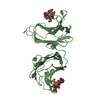
| 登録情報 | データベース: PDB / ID: 1qg5 | ||||||
|---|---|---|---|---|---|---|---|
| タイトル | HIGH RESOLUTION CRYSTAL STRUCTURE OF THE BOVINE BETA-LACTOGLOBULIN (ISOFORM A) | ||||||
 要素 要素 | BETA-LACTOGLOBULIN | ||||||
 キーワード キーワード | TRANSPORT PROTEIN / LIPOCALIN / ISOFORM A | ||||||
| 機能・相同性 |  機能・相同性情報 機能・相同性情報retinol binding / long-chain fatty acid binding / extracellular region / identical protein binding 類似検索 - 分子機能 | ||||||
| 生物種 |  | ||||||
| 手法 |  X線回折 / シンクロトロン / 分子置換 / 解像度: 2 Å X線回折 / シンクロトロン / 分子置換 / 解像度: 2 Å | ||||||
 データ登録者 データ登録者 | Oliveira, K.M.G. / Sawyer, L. / Polikarpov, I. | ||||||
 引用 引用 | ジャーナル: Eur.J.Biochem. / 年: 2001 タイトル: Crystal structures of bovine beta-lactoglobulin in the orthorhombic space group C222(1). Structural differences between genetic variants A and B and features of the Tanford transition. 著者: Oliveira, K.M. / Valente-Mesquita, V.L. / Botelho, M.M. / Sawyer, L. / Ferreira, S.T. / Polikarpov, I. | ||||||
| 履歴 |
|
- 構造の表示
構造の表示
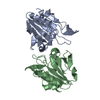
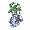
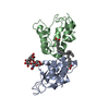
| 構造ビューア | 分子: MolmilJmol/JSmol |
|---|
- ダウンロードとリンク
-ダウンロード
| PDBx/mmCIF形式 | 1qg5.cif.gz | 45 KB | 表示 | PDBx/mmCIF形式 |
|---|---|---|---|---|
| PDB形式 | pdb1qg5.ent.gz | 31.6 KB | 表示 | PDB形式 |
| PDBx/mmJSON形式 | 1qg5.json.gz | ツリー表示 | PDBx/mmJSON形式 | |
| その他 |  その他のダウンロード その他のダウンロード |
-検証レポート
| アーカイブディレクトリ | https://data.pdbj.org/pub/pdb/validation_reports/qg/1qg5ftp://data.pdbj.org/pub/pdb/validation_reports/qg/1qg5 | HTTPS FTP |
|---|
-関連構造データ


-リンク
 PDBj
PDBj

- 集合体
集合体
| 登録構造単位 | 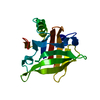
| ||||||||||
|---|---|---|---|---|---|---|---|---|---|---|---|
| 1 | 
| ||||||||||
| 単位格子 |
| ||||||||||
| Components on special symmetry positions |
|
-要素
| #1: タンパク質 | 分子量: 18387.264 Da / 分子数: 1 / 由来タイプ: 天然 詳細: ISOFORM A DIFFERS IN A PRIMARY AMINO ACID SEQUENCE FROM ISOFORM B AT POSITIONS 64(ASP-->GLY) AND 118 (VAL-->ALA) 由来: (天然) |
|---|---|
| #2: 水 | ChemComp-HOH /  分子量: 18.015 Da / 分子数: 113 / 由来タイプ: 天然 / 式: H2O 分子量: 18.015 Da / 分子数: 113 / 由来タイプ: 天然 / 式: H2O |
| Has protein modification | Y |
-実験情報
-実験
| 実験 | 手法: X線回折 / 使用した結晶の数: 1 |
|---|
- 試料調製
試料調製
| 結晶 | マシュー密度: 2.1 Å3/Da / 溶媒含有率: 42 % / 解説: DATA WERE COLLECTED USING OSCILLATION METHOD | ||||||||||||||||||||
|---|---|---|---|---|---|---|---|---|---|---|---|---|---|---|---|---|---|---|---|---|---|
| 結晶化 | 手法: 蒸気拡散法, ハンギングドロップ法 / pH: 7.9 詳細: THE CRYSTAL WAS GROWN AT PH 7.9, HANGING DROP METHOD. DROP CONTAINED 2.5M (NH4) 2SO4, 180MM TRIZMA BUFFER, AND 20MG/ML PROTEIN REMARK 290, VAPOR DIFFUSION, HANGING DROP | ||||||||||||||||||||
| 結晶化 | *PLUS | ||||||||||||||||||||
| 溶液の組成 | *PLUS
|
-データ収集
| 回折 | 平均測定温度: 291 K |
|---|---|
| 放射光源 | 由来: シンクロトロン / サイト: LNLS  / ビームライン: D03B-MX1 / 波長: 1.38 / ビームライン: D03B-MX1 / 波長: 1.38 |
| 検出器 | タイプ: MARRESEARCH / 検出器: IMAGE PLATE / 日付: 1997年9月15日 / 詳細: MIRRORS |
| 放射 | モノクロメーター: SI (111) / プロトコル: SINGLE WAVELENGTH / 単色(M)・ラウエ(L): M / 散乱光タイプ: x-ray |
| 放射波長 | 波長: 1.38 Å / 相対比: 1 |
| 反射 | 解像度: 2→12.92 Å / Num. obs: 9631 / % possible obs: 90.9 % / Observed criterion σ(I): 2 / 冗長度: 2.8 % / Biso Wilson estimate: 28.4 Å2 / Rsym value: 0.066 / Net I/σ(I): 12.7 |
| 反射 シェル | 解像度: 2→2.05 Å / Rsym value: 0.463 / % possible all: 91.2 |
| 反射 | *PLUS 最低解像度: 12.9 Å / Rmerge(I) obs: 0.066 |
| 反射 シェル | *PLUS % possible obs: 91.2 % / Rmerge(I) obs: 0.46 / Mean I/σ(I) obs: 2.6 |
- 解析
解析
| ソフトウェア |
| |||||||||||||||||||||||||||||||||||||||||||||||||||||||||||||||
|---|---|---|---|---|---|---|---|---|---|---|---|---|---|---|---|---|---|---|---|---|---|---|---|---|---|---|---|---|---|---|---|---|---|---|---|---|---|---|---|---|---|---|---|---|---|---|---|---|---|---|---|---|---|---|---|---|---|---|---|---|---|---|---|---|
| 精密化 | 構造決定の手法: 分子置換 / 解像度: 2→9.5 Å / 交差検証法: THROUGHOUT / σ(F): 0 / ESU R: 0.21
| |||||||||||||||||||||||||||||||||||||||||||||||||||||||||||||||
| 原子変位パラメータ | Biso mean: 42.9 Å2 | |||||||||||||||||||||||||||||||||||||||||||||||||||||||||||||||
| 精密化ステップ | サイクル: LAST / 解像度: 2→9.5 Å
| |||||||||||||||||||||||||||||||||||||||||||||||||||||||||||||||
| 拘束条件 |
| |||||||||||||||||||||||||||||||||||||||||||||||||||||||||||||||
| ソフトウェア | *PLUS 名称: REFMAC / 分類: refinement | |||||||||||||||||||||||||||||||||||||||||||||||||||||||||||||||
| 精密化 | *PLUS Rfactor Rfree: 0.283 / Rfactor Rwork: 0.212 | |||||||||||||||||||||||||||||||||||||||||||||||||||||||||||||||
| 溶媒の処理 | *PLUS | |||||||||||||||||||||||||||||||||||||||||||||||||||||||||||||||
| 原子変位パラメータ | *PLUS | |||||||||||||||||||||||||||||||||||||||||||||||||||||||||||||||
| 拘束条件 | *PLUS
|
