 Movie
Movie Controller

[English] 日本語
 Yorodumi
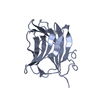
- PDB-1w8w: CBM29-2 mutant Y46A: Probing the Mechanism of Ligand Recognition ... -
+ Open data

- Basic information
| Entry | Database: PDB / ID: 1w8w | ||||||
|---|---|---|---|---|---|---|---|
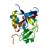
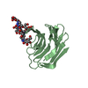
| Title | CBM29-2 mutant Y46A: Probing the Mechanism of Ligand Recognition by Family 29 Carbohydrate Binding Modules | ||||||
 Components Components | NON-CATALYTIC PROTEIN 1 | ||||||
 Keywords Keywords | CARBOHYDRATE BINDING DOMAIN / CARBOHYDRATE BINDING MODULE / GLUCOMANNAN / CELLOHEXAOSE / MANNOHEXAOSE / CELLULOSOME | ||||||
| Function / homology |  Function and homology information Function and homology information | ||||||
| Biological species |  PIROMYCES EQUI (fungus) PIROMYCES EQUI (fungus) | ||||||
| Method |  X-RAY DIFFRACTION / SYNCHROTRON / MOLECULAR REPLACEMENT / Resolution: 2.1 Å X-RAY DIFFRACTION / SYNCHROTRON / MOLECULAR REPLACEMENT / Resolution: 2.1 Å | ||||||
 Authors Authors | Flint, J. / Bolam, D.N. / Nurizzo, D. / Taylor, E.J. / Williamson, M.P. / Walters, C. / Davies, G.J. / Gilbert, H.J. | ||||||
 Citation Citation | Journal: J.Biol.Chem. / Year: 2005 Title: Probing the Mechanism of Ligand Recognition in Family 29 Carbohydrate-Binding Modules Authors: Flint, J. / Bolam, D.N. / Nurizzo, D. / Taylor, E.J. / Williamson, M.P. / Walters, C. / Davies, G.J. / Gilbert, H.J. | ||||||
| History |
| ||||||
| Remark 700 | SHEET THE SHEET STRUCTURE OF THIS MOLECULE IS BIFURCATED. IN ORDER TO REPRESENT THIS FEATURE IN ... SHEET THE SHEET STRUCTURE OF THIS MOLECULE IS BIFURCATED. IN ORDER TO REPRESENT THIS FEATURE IN THE SHEET RECORDS BELOW, TWO SHEETS ARE DEFINED. |
- Structure visualization
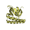
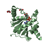
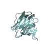
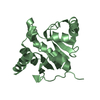
| Structure viewer | Molecule: MolmilJmol/JSmol |
|---|
- Downloads & links
-Download
| PDBx/mmCIF format | 1w8w.cif.gz | 69.3 KB | Display | PDBx/mmCIF format |
|---|---|---|---|---|
| PDB format | pdb1w8w.ent.gz | 52.2 KB | Display | PDB format |
| PDBx/mmJSON format | 1w8w.json.gz | Tree view | PDBx/mmJSON format | |
| Others |  Other downloads Other downloads |
-Validation report
| Arichive directory | https://data.pdbj.org/pub/pdb/validation_reports/w8/1w8wftp://data.pdbj.org/pub/pdb/validation_reports/w8/1w8w | HTTPS FTP |
|---|
-Related structure data
| Related structure data |  1w8tC  1w8uC  1w8zC  1w90C  1w9fC  1wcuC  1gwmS S: Starting model for refinement C: citing same article ( |
|---|---|
| Similar structure data |


-Links
 PDBj
PDBj


- Assembly
| Deposited unit | 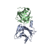
| ||||||||
|---|---|---|---|---|---|---|---|---|---|
| 1 | 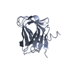
| ||||||||
| 2 | 
| ||||||||
| Unit cell |
| ||||||||
| Components on special symmetry positions |
|
-Components
| #1: Protein | Mass: 16574.250 Da / Num. of mol.: 2 / Fragment: CARBOHYDRATE BINDING MODULE 2, RESIDUES 334-478 / Mutation: YES Source method: isolated from a genetically manipulated source Source: (gene. exp.) PIROMYCES EQUI (fungus) / Plasmid: PET22B / Production host:  #2: Water | ChemComp-HOH / |  Mass: 18.015 Da / Num. of mol.: 89 / Source method: isolated from a natural source / Formula: H2O Mass: 18.015 Da / Num. of mol.: 89 / Source method: isolated from a natural source / Formula: H2OCompound details | ENGINEERED | |
|---|
-Experimental details
-Experiment
| Experiment | Method: X-RAY DIFFRACTION / Number of used crystals: 1 |
|---|
- Sample preparation
| Crystal | Density Matthews: 3.1 Å3/Da / Density % sol: 60 % |
|---|---|
| Crystal grow | pH: 7.5 Details: 100 MM NA/HEPES BUFFER PH 7.5, CONTAINING 150 MM KSCN, 20% ETHYLENE GLYCOL 20%PEG3350 |
-Data collection
| Diffraction | Mean temperature: 100 K |
|---|---|
| Diffraction source | Source: SYNCHROTRON / Site: ESRF  / Beamline: ID14-1 / Wavelength: 0.934 / Beamline: ID14-1 / Wavelength: 0.934 |
| Radiation | Protocol: SINGLE WAVELENGTH / Monochromatic (M) / Laue (L): M / Scattering type: x-ray |
| Radiation wavelength | Wavelength: 0.934 Å / Relative weight: 1 |
| Reflection | Resolution: 2.1→20 Å / Num. obs: 21019 / % possible obs: 99.9 % / Redundancy: 7.3 % / Rmerge(I) obs: 0.08 / Net I/σ(I): 21.8 |
| Reflection shell | Resolution: 2.1→2.21 Å / Redundancy: 7.5 % / Rmerge(I) obs: 0.41 / Mean I/σ(I) obs: 4.3 / % possible all: 100 |
- Processing
| Software |
| ||||||||||||||||||||||||||||||||||||||||||||||||||||||||||||||||||||||||||||||||||||||||||||||||||||||||||||||||||||||||||||||||||||||||||||||||||||||||||||||||||||||||||||||||||||||
|---|---|---|---|---|---|---|---|---|---|---|---|---|---|---|---|---|---|---|---|---|---|---|---|---|---|---|---|---|---|---|---|---|---|---|---|---|---|---|---|---|---|---|---|---|---|---|---|---|---|---|---|---|---|---|---|---|---|---|---|---|---|---|---|---|---|---|---|---|---|---|---|---|---|---|---|---|---|---|---|---|---|---|---|---|---|---|---|---|---|---|---|---|---|---|---|---|---|---|---|---|---|---|---|---|---|---|---|---|---|---|---|---|---|---|---|---|---|---|---|---|---|---|---|---|---|---|---|---|---|---|---|---|---|---|---|---|---|---|---|---|---|---|---|---|---|---|---|---|---|---|---|---|---|---|---|---|---|---|---|---|---|---|---|---|---|---|---|---|---|---|---|---|---|---|---|---|---|---|---|---|---|---|---|
| Refinement | Method to determine structure: MOLECULAR REPLACEMENT Starting model: PDB ENTRY 1GWM Resolution: 2.1→65.94 Å / Cor.coef. Fo:Fc: 0.95 / Cor.coef. Fo:Fc free: 0.939 / SU B: 4.405 / SU ML: 0.116 / TLS residual ADP flag: LIKELY RESIDUAL / Cross valid method: THROUGHOUT / ESU R: 0.2 / ESU R Free: 0.171 / Stereochemistry target values: MAXIMUM LIKELIHOOD / Details: HYDROGENS HAVE BEEN ADDED IN THE RIDING POSITIONS.
| ||||||||||||||||||||||||||||||||||||||||||||||||||||||||||||||||||||||||||||||||||||||||||||||||||||||||||||||||||||||||||||||||||||||||||||||||||||||||||||||||||||||||||||||||||||||
| Solvent computation | Ion probe radii: 0.8 Å / Shrinkage radii: 0.8 Å / VDW probe radii: 1.4 Å / Solvent model: BABINET MODEL WITH MASK | ||||||||||||||||||||||||||||||||||||||||||||||||||||||||||||||||||||||||||||||||||||||||||||||||||||||||||||||||||||||||||||||||||||||||||||||||||||||||||||||||||||||||||||||||||||||
| Displacement parameters | Biso mean: 19.63 Å2
| ||||||||||||||||||||||||||||||||||||||||||||||||||||||||||||||||||||||||||||||||||||||||||||||||||||||||||||||||||||||||||||||||||||||||||||||||||||||||||||||||||||||||||||||||||||||
| Refinement step | Cycle: LAST / Resolution: 2.1→65.94 Å
| ||||||||||||||||||||||||||||||||||||||||||||||||||||||||||||||||||||||||||||||||||||||||||||||||||||||||||||||||||||||||||||||||||||||||||||||||||||||||||||||||||||||||||||||||||||||
| Refine LS restraints |
|