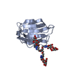
Resolution: 1.79→45.495 Å / SU ML: 0.21 / σ(F): 1.35 / Phase error: 27.08 / Stereochemistry target values: ML Details: RESIDUES 1-4 ARE DISORDERED ELECTRON DENSITY FOR ONLY FOUR OF SIX ARMS FOR THE DENDRON IS OBSERVED. PDB FILE CONTAINS ONLY THESE FOUR ARMS.
Rfactor
Num. reflection
% reflection
Rfree
0.2509
1247
5.1 %
Rwork
0.2002
-
-
obs
0.2028
24359
95.11 %
Solvent computation
Shrinkage radii: 0.9 Å / VDW probe radii: 1.11 Å / Solvent model: FLAT BULK SOLVENT MODEL
Refinement step
Cycle: LAST / Resolution: 1.79→45.495 Å
Protein
Nucleic acid
Ligand
Solvent
Total
Num. atoms
2067
0
95
156
2318
Refine LS restraints
Refine-ID
Type
Dev ideal
Number
X-RAY DIFFRACTION
f_bond_d
0.012
2249
X-RAY DIFFRACTION
f_angle_d
1.437
3059
X-RAY DIFFRACTION
f_dihedral_angle_d
15.538
965
X-RAY DIFFRACTION
f_chiral_restr
0.147
322
X-RAY DIFFRACTION
f_plane_restr
0.007
408
LS refinement shell
Resolution (Å)
Rfactor Rfree
Num. reflection Rfree
Rfactor Rwork
Num. reflection Rwork
Refine-ID
% reflection obs (%)
1.79-1.8617
0.3064
130
0.2443
2432
X-RAY DIFFRACTION
91
1.8617-1.9464
0.2511
169
0.2143
2535
X-RAY DIFFRACTION
95
1.9464-2.0491
0.2681
135
0.2086
2583
X-RAY DIFFRACTION
96
2.0491-2.1774
0.2959
126
0.2044
2590
X-RAY DIFFRACTION
96
2.1774-2.3455
0.2556
125
0.2137
2510
X-RAY DIFFRACTION
93
2.3455-2.5816
0.2351
119
0.2131
2683
X-RAY DIFFRACTION
98
2.5816-2.9551
0.2915
139
0.2291
2607
X-RAY DIFFRACTION
97
2.9551-3.7228
0.25
136
0.1926
2559
X-RAY DIFFRACTION
94
3.7228-45.5093
0.22
168
0.171
2613
X-RAY DIFFRACTION
95
+
About Yorodumi
-
News
-
Feb 9, 2022. New format data for meta-information of EMDB entries
New format data for meta-information of EMDB entries
Version 3 of the EMDB header file is now the official format.
The previous official version 1.9 will be removed from the archive.
In the structure databanks used in Yorodumi, some data are registered as the other names, "COVID-19 virus" and "2019-nCoV". Here are the details of the virus and the list of structure data.
Jan 31, 2019. EMDB accession codes are about to change! (news from PDBe EMDB page)
EMDB accession codes are about to change! (news from PDBe EMDB page)
The allocation of 4 digits for EMDB accession codes will soon come to an end. Whilst these codes will remain in use, new EMDB accession codes will include an additional digit and will expand incrementally as the available range of codes is exhausted. The current 4-digit format prefixed with “EMD-” (i.e. EMD-XXXX) will advance to a 5-digit format (i.e. EMD-XXXXX), and so on. It is currently estimated that the 4-digit codes will be depleted around Spring 2019, at which point the 5-digit format will come into force.
The EM Navigator/Yorodumi systems omit the EMD- prefix.
Related info.:Q: What is EMD? / ID/Accession-code notation in Yorodumi/EM Navigator
Yorodumi is a browser for structure data from EMDB, PDB, SASBDB, etc.
This page is also the successor to EM Navigator detail page, and also detail information page/front-end page for Omokage search.
The word "yorodu" (or yorozu) is an old Japanese word meaning "ten thousand". "mi" (miru) is to see.
Related info.:EMDB / PDB / SASBDB / Comparison of 3 databanks / Yorodumi Search / Aug 31, 2016. New EM Navigator & Yorodumi / Yorodumi Papers / Jmol/JSmol / Function and homology information / Changes in new EM Navigator and Yorodumi
 Movie
Movie Controller


 Open data
 Basic information
 Components
 Keywords
 Function and homology information
 HOMO SAPIENS (human)
 X-RAY DIFFRACTION /
  Authors
 Citation
 Structure visualization
 Downloads & links
 Other downloads
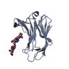


 PDBj
PDBj Assembly


 Mass: 1810.460 Da / Num. of mol.: 1 / Source method: obtained synthetically / Formula: C69H77N20O39
 Mass: 18.015 Da / Num. of mol.: 156 / Source method: isolated from a natural source / Formula: H2O
 Sample preparation
 / Beamline: I04-1 / Wavelength: 0.92
  Processing