 Movie
Movie Controller


+ Open data

- Basic information
| Entry | Database: PDB / ID: 4uw5 | ||||||
|---|---|---|---|---|---|---|---|
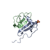
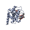
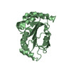
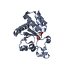
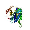
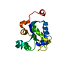
| Title | Human galectin-7 in complex with a galactose based dendron D2-2. | ||||||
 Components Components | HUMAN GALECTIN-7 | ||||||
 Keywords Keywords | SUGAR BINDING PROTEIN / LECTIN / DENDRIMERS / MULTIVALENCY / CARBOHYDRATE BINDING | ||||||
| Function / homology |  Function and homology information Function and homology informationDifferentiation of Keratinocytes in Interfollicular Epidermis in Mammalian Skin / heterophilic cell-cell adhesion / carbohydrate binding / apoptotic process / : / extracellular exosome / nucleus / cytoplasm Similarity search - Function | ||||||
| Biological species |  HOMO SAPIENS (human) HOMO SAPIENS (human) | ||||||
| Method |  X-RAY DIFFRACTION / SYNCHROTRON / MOLECULAR REPLACEMENT / Resolution: 2.04 Å X-RAY DIFFRACTION / SYNCHROTRON / MOLECULAR REPLACEMENT / Resolution: 2.04 Å | ||||||
 Authors Authors | Ramaswamy, S. / Sleiman, M.H. / Masuyer, G. / Arbez-Gindre, C. / Micha-Screttas, M. / Calogeropoulou, T. / Steele, B.R. / Acharya, K.R. | ||||||
 Citation Citation | Journal: FEBS J. / Year: 2015 Title: Structural Basis of Multivalent Galactose-Based Dendrimer Recognition by Human Galectin-7. Authors: Ramaswamy, S. / Haj Sleiman, M. / Masuyer, G. / Arbez-Gindre, C. / Micha-Screttas, M. / Calogeropoulou, T. / Steele, B.R. / Acharya, K.R. | ||||||
| History |
|
- Structure visualization
| Structure viewer | Molecule: MolmilJmol/JSmol |
|---|
- Downloads & links
-Download
| PDBx/mmCIF format | 4uw5.cif.gz | 176.9 KB | Display | PDBx/mmCIF format |
|---|---|---|---|---|
| PDB format | pdb4uw5.ent.gz | 143.4 KB | Display | PDB format |
| PDBx/mmJSON format | 4uw5.json.gz | Tree view | PDBx/mmJSON format | |
| Others |  Other downloads Other downloads |
-Validation report
| Arichive directory | https://data.pdbj.org/pub/pdb/validation_reports/uw/4uw5ftp://data.pdbj.org/pub/pdb/validation_reports/uw/4uw5 | HTTPS FTP |
|---|
-Related structure data
| Related structure data | 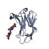 4uw3C 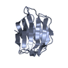 4uw4C  4uw6C 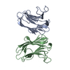 1bkzS C: citing same article ( S: Starting model for refinement |
|---|---|
| Similar structure data |


-Links
 PDBj
PDBj- Assembly
| Deposited unit | 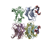
| ||||||||
|---|---|---|---|---|---|---|---|---|---|
| 1 | 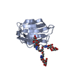
| ||||||||
| 2 | 
| ||||||||
| 3 | 
| ||||||||
| 4 | 
| ||||||||
| 5 | 
| ||||||||
| 6 | 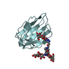
| ||||||||
| Unit cell |
|
-Components
| #1: Protein | Mass: 15097.046 Da / Num. of mol.: 6 Source method: isolated from a genetically manipulated source Source: (gene. exp.) HOMO SAPIENS (human) / Plasmid: PET-22 / Production host:  #2: Chemical |   Mass: 1337.207 Da / Num. of mol.: 2 / Source method: obtained synthetically / Formula: C49H80N10O33 Mass: 1337.207 Da / Num. of mol.: 2 / Source method: obtained synthetically / Formula: C49H80N10O33#3: Water | ChemComp-HOH / |  Mass: 18.015 Da / Num. of mol.: 422 / Source method: isolated from a natural source / Formula: H2O Mass: 18.015 Da / Num. of mol.: 422 / Source method: isolated from a natural source / Formula: H2O |
|---|
-Experimental details
-Experiment
| Experiment | Method: X-RAY DIFFRACTION / Number of used crystals: 1 |
|---|
- Sample preparation
| Crystal | Density Matthews: 2.22 Å3/Da / Density % sol: 44.6 % / Description: NONE |
|---|---|
| Crystal grow | Details: 0.05M BIS TRIS PROPANE PH7, 14% PEG 3350 |
-Data collection
| Diffraction | Mean temperature: 100 K |
|---|---|
| Diffraction source | Source: SYNCHROTRON / Site: Diamond  / Beamline: I03 / Wavelength: 0.9783 / Beamline: I03 / Wavelength: 0.9783 |
| Detector | Type: DECTRIS PILATUS 6M / Detector: PIXEL / Date: Jul 8, 2013 |
| Radiation | Protocol: SINGLE WAVELENGTH / Monochromatic (M) / Laue (L): M / Scattering type: x-ray |
| Radiation wavelength | Wavelength: 0.9783 Å / Relative weight: 1 |
| Reflection | Resolution: 2.04→55.69 Å / Num. obs: 54509 / % possible obs: 98.7 % / Observed criterion σ(I): 0 / Redundancy: 3.3 % / Biso Wilson estimate: 32.54 Å2 / Rmerge(I) obs: 0.07 / Net I/σ(I): 9.1 |
| Reflection shell | Resolution: 2.04→2.11 Å / Redundancy: 3.3 % / Rmerge(I) obs: 0.6 / Mean I/σ(I) obs: 2.1 / % possible all: 99.3 |
- Processing
| Software |
| |||||||||||||||||||||||||||||||||||||||||||||||||||||||||||||||||||||||||||||
|---|---|---|---|---|---|---|---|---|---|---|---|---|---|---|---|---|---|---|---|---|---|---|---|---|---|---|---|---|---|---|---|---|---|---|---|---|---|---|---|---|---|---|---|---|---|---|---|---|---|---|---|---|---|---|---|---|---|---|---|---|---|---|---|---|---|---|---|---|---|---|---|---|---|---|---|---|---|---|
| Refinement | Method to determine structure: MOLECULAR REPLACEMENT Starting model: PDB ENTRY 1BKZ Resolution: 2.04→55.687 Å / SU ML: 0.35 / σ(F): 1.35 / Phase error: 36.18 / Stereochemistry target values: ML Details: RESIDUES 1-3 ARE DISORDERED DENSITY MODIFICATION PERFORMED. MTZ FILE POST DENSITY MODIFICATION SUBMITTED.
| |||||||||||||||||||||||||||||||||||||||||||||||||||||||||||||||||||||||||||||
| Solvent computation | Shrinkage radii: 0.9 Å / VDW probe radii: 1.11 Å / Solvent model: FLAT BULK SOLVENT MODEL | |||||||||||||||||||||||||||||||||||||||||||||||||||||||||||||||||||||||||||||
| Refinement step | Cycle: LAST / Resolution: 2.04→55.687 Å
| |||||||||||||||||||||||||||||||||||||||||||||||||||||||||||||||||||||||||||||
| Refine LS restraints |
| |||||||||||||||||||||||||||||||||||||||||||||||||||||||||||||||||||||||||||||
| LS refinement shell |
|
