 Movie
Movie Controller

[English] 日本語
 Yorodumi
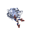
- PDB-3wde: Mutant N-terminal domain of Mycobacterium tuberculosis ClpC1, F80... -
+ Open data

- Basic information
| Entry | Database: PDB / ID: 3wde | ||||||
|---|---|---|---|---|---|---|---|
| Title | Mutant N-terminal domain of Mycobacterium tuberculosis ClpC1, F80Y, bound to Cyclomarin A | ||||||
 Components Components |
| ||||||
 Keywords Keywords | Chaperone/Antimicrobial protein / Chaperone / Chaperone-Antimicrobial protein complex | ||||||
| Function / homology |  Function and homology information Function and homology informationprotein folding chaperone / peptidoglycan-based cell wall / protein homodimerization activity / ATP hydrolysis activity / ATP binding / plasma membrane / cytosol Similarity search - Function | ||||||
| Biological species |   Mycobacterium tuberculosis (bacteria)Streptomyces (bacteria) Mycobacterium tuberculosis (bacteria)Streptomyces (bacteria) | ||||||
| Method |  X-RAY DIFFRACTION / SYNCHROTRON / MOLECULAR REPLACEMENT / Resolution: 1.44 Å X-RAY DIFFRACTION / SYNCHROTRON / MOLECULAR REPLACEMENT / Resolution: 1.44 Å | ||||||
 Authors Authors | Vasudevan, D. / Noble, C.G. | ||||||
 Citation Citation | Journal: J.Biol.Chem. / Year: 2013 Title: Structural basis of mycobacterial inhibition by cyclomarin A Authors: Vasudevan, D. / Rao, S.P.S. / Noble, C.G. | ||||||
| History |
|
- Structure visualization
| Structure viewer | Molecule: MolmilJmol/JSmol |
|---|
- Downloads & links
-Download
| PDBx/mmCIF format | 3wde.cif.gz | 86.7 KB | Display | PDBx/mmCIF format |
|---|---|---|---|---|
| PDB format | pdb3wde.ent.gz | 65.2 KB | Display | PDB format |
| PDBx/mmJSON format | 3wde.json.gz | Tree view | PDBx/mmJSON format | |
| Others |  Other downloads Other downloads |
-Validation report
| Arichive directory | https://data.pdbj.org/pub/pdb/validation_reports/wd/3wdeftp://data.pdbj.org/pub/pdb/validation_reports/wd/3wde | HTTPS FTP |
|---|
-Related structure data
| Related structure data |  3wdbC 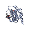 3wdcSC  3wddC C: citing same article ( S: Starting model for refinement |
|---|---|
| Similar structure data |


-Links
 PDBj
PDBj


- Assembly
| Deposited unit | 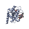
| ||||||||
|---|---|---|---|---|---|---|---|---|---|
| 1 |
| ||||||||
| Unit cell |
|
-Components
| #1: Protein | Mass: 17089.557 Da / Num. of mol.: 1 / Fragment: N-terminal domain, UNP residues 1-145 / Mutation: F80Y Source method: isolated from a genetically manipulated source Source: (gene. exp.) Mycobacterium tuberculosis (bacteria) / Gene: clpC / Production host: |
|---|---|
| #2: Protein/peptide | 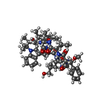  Type: Cyclic peptide / Class: Antiinflammatory / Mass: 1063.328 Da / Num. of mol.: 1 / Source method: isolated from a natural source / Source: (natural) Streptomyces (bacteria) / References: Cyclomarin A Type: Cyclic peptide / Class: Antiinflammatory / Mass: 1063.328 Da / Num. of mol.: 1 / Source method: isolated from a natural source / Source: (natural) Streptomyces (bacteria) / References: Cyclomarin A |
| #3: Chemical | ChemComp-ACT /   Mass: 59.044 Da / Num. of mol.: 1 / Source method: obtained synthetically / Formula: C2H3O2 Mass: 59.044 Da / Num. of mol.: 1 / Source method: obtained synthetically / Formula: C2H3O2 |
| #4: Water | ChemComp-HOH /  Mass: 18.015 Da / Num. of mol.: 160 / Source method: isolated from a natural source / Formula: H2O Mass: 18.015 Da / Num. of mol.: 160 / Source method: isolated from a natural source / Formula: H2O |
| Has protein modification | Y |
-Experimental details
-Experiment
| Experiment | Method: X-RAY DIFFRACTION / Number of used crystals: 1 |
|---|
- Sample preparation
| Crystal | Density Matthews: 1.76 Å3/Da / Density % sol: 30.3 % |
|---|---|
| Crystal grow | Temperature: 291 K / Method: vapor diffusion, hanging drop / pH: 7.5 Details: 0.2M ammonium acetate, 20% polyethylene glycol 3350, 0.025M magnesium formate, pH 7.5, VAPOR DIFFUSION, HANGING DROP, temperature 291K |
-Data collection
| Diffraction | Mean temperature: 298 K |
|---|---|
| Diffraction source | Source: SYNCHROTRON / Site: SLS  / Beamline: X10SA / Wavelength: 1 Å / Beamline: X10SA / Wavelength: 1 Å |
| Detector | Type: PSI PILATUS 6M / Detector: PIXEL / Date: Feb 1, 2011 |
| Radiation | Protocol: SINGLE WAVELENGTH / Monochromatic (M) / Laue (L): M / Scattering type: x-ray |
| Radiation wavelength | Wavelength: 1 Å / Relative weight: 1 |
| Reflection | Resolution: 1.44→50 Å / Num. obs: 23876 |
- Processing
| Software |
| ||||||||||||||||||||||||||||||||||||||||||||||||||||||||||||
|---|---|---|---|---|---|---|---|---|---|---|---|---|---|---|---|---|---|---|---|---|---|---|---|---|---|---|---|---|---|---|---|---|---|---|---|---|---|---|---|---|---|---|---|---|---|---|---|---|---|---|---|---|---|---|---|---|---|---|---|---|---|
| Refinement | Method to determine structure: MOLECULAR REPLACEMENT Starting model: 3WDC Resolution: 1.44→30 Å / Cor.coef. Fo:Fc: 0.977 / Cor.coef. Fo:Fc free: 0.962 / Occupancy max: 1 / Occupancy min: 0.2 / SU B: 1.894 / SU ML: 0.034 / Cross valid method: THROUGHOUT / ESU R: 0.075 / ESU R Free: 0.067 / Stereochemistry target values: MAXIMUM LIKELIHOOD Details: HYDROGENS HAVE BEEN USED IF PRESENT IN THE INPUT U VALUES: REFINED INDIVIDUALLY
| ||||||||||||||||||||||||||||||||||||||||||||||||||||||||||||
| Solvent computation | Ion probe radii: 0.8 Å / Shrinkage radii: 0.8 Å / VDW probe radii: 1.2 Å / Solvent model: MASK | ||||||||||||||||||||||||||||||||||||||||||||||||||||||||||||
| Displacement parameters | Biso max: 177.93 Å2 / Biso mean: 13.8438 Å2 / Biso min: 3.47 Å2
| ||||||||||||||||||||||||||||||||||||||||||||||||||||||||||||
| Refinement step | Cycle: LAST / Resolution: 1.44→30 Å
| ||||||||||||||||||||||||||||||||||||||||||||||||||||||||||||
| Refine LS restraints |
| ||||||||||||||||||||||||||||||||||||||||||||||||||||||||||||
| LS refinement shell | Resolution: 1.44→1.477 Å / Total num. of bins used: 20
|
