 Movie
Movie Controller

[English] 日本語
 Yorodumi
- PDB-1npk: REFINED X-RAY STRUCTURE OF DICTYOSTELIUM NUCLEOSIDE DIPHOSPHATE K... -
+ Open data

- Basic information
| Entry | Database: PDB / ID: 1npk | ||||||
|---|---|---|---|---|---|---|---|
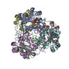
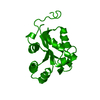
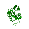
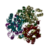
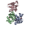
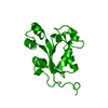
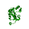
| Title | REFINED X-RAY STRUCTURE OF DICTYOSTELIUM NUCLEOSIDE DIPHOSPHATE KINASE AT 1,8 ANGSTROMS RESOLUTION | ||||||
 Components Components | NUCLEOSIDE DIPHOSPHATE KINASE | ||||||
 Keywords Keywords | PHOSPHOTRANSFERASE(PO4 AS ACCEPTOR) | ||||||
| Function / homology |  Function and homology information Function and homology information: / Azathioprine ADME / Ribavirin ADME / asexual reproduction / Interconversion of nucleotide di- and triphosphates / Neutrophil degranulation / negative regulation of pinocytosis / nucleoside triphosphate biosynthetic process / nucleoside-diphosphate kinase / UTP biosynthetic process ...: / Azathioprine ADME / Ribavirin ADME / asexual reproduction / Interconversion of nucleotide di- and triphosphates / Neutrophil degranulation / negative regulation of pinocytosis / nucleoside triphosphate biosynthetic process / nucleoside-diphosphate kinase / UTP biosynthetic process / CTP biosynthetic process / nucleoside diphosphate kinase activity / GTP biosynthetic process / negative regulation of exocytosis / negative regulation of phagocytosis / translational elongation / phagocytic vesicle / secretory granule / response to bacterium / actin cytoskeleton organization / cytoskeleton / ribosome / G protein-coupled receptor signaling pathway / ATP binding / metal ion binding / plasma membrane / cytoplasm Similarity search - Function | ||||||
| Biological species |  | ||||||
| Method |  X-RAY DIFFRACTION / Resolution: 1.8 Å X-RAY DIFFRACTION / Resolution: 1.8 Å | ||||||
 Authors Authors | Janin, J. / Morera, S. / Lebras, G. / Lascu, I. / Veron, M. | ||||||
 Citation Citation | Journal: J.Mol.Biol. / Year: 1994 Title: Refined X-ray structure of Dictyostelium discoideum nucleoside diphosphate kinase at 1.8 A resolution. Authors: Morera, S. / LeBras, G. / Lascu, I. / Lacombe, M.L. / Veron, M. / Janin, J. | ||||||
| History |
|
- Structure visualization
| Structure viewer | Molecule: MolmilJmol/JSmol |
|---|
- Downloads & links
-Download
| PDBx/mmCIF format | 1npk.cif.gz | 41.2 KB | Display | PDBx/mmCIF format |
|---|---|---|---|---|
| PDB format | pdb1npk.ent.gz | 29.2 KB | Display | PDB format |
| PDBx/mmJSON format | 1npk.json.gz | Tree view | PDBx/mmJSON format | |
| Others |  Other downloads Other downloads |
-Validation report
| Arichive directory | https://data.pdbj.org/pub/pdb/validation_reports/np/1npkftp://data.pdbj.org/pub/pdb/validation_reports/np/1npk | HTTPS FTP |
|---|
-Related structure data
| Similar structure data |
|---|


-Links
 PDBj
PDBj
- Assembly
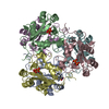
| Deposited unit | 
| ||||||||
|---|---|---|---|---|---|---|---|---|---|
| 1 | x 6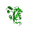
| ||||||||
| Unit cell |
|
-Components
| #1: Protein | Mass: 16685.139 Da / Num. of mol.: 1 Source method: isolated from a genetically manipulated source Source: (gene. exp.) |
|---|---|
| #2: Water | ChemComp-HOH /  Mass: 18.015 Da / Num. of mol.: 95 / Source method: isolated from a natural source / Formula: H2O Mass: 18.015 Da / Num. of mol.: 95 / Source method: isolated from a natural source / Formula: H2O |
-Experimental details
-Experiment
| Experiment | Method: X-RAY DIFFRACTION |
|---|
- Sample preparation
| Crystal | Density Matthews: 2.5 Å3/Da / Density % sol: 50.85 % | ||||||||||||||||||||||||||||||||||||
|---|---|---|---|---|---|---|---|---|---|---|---|---|---|---|---|---|---|---|---|---|---|---|---|---|---|---|---|---|---|---|---|---|---|---|---|---|---|
| Crystal grow | *PLUS Temperature: 18 ℃ / pH: 8 / Method: vapor diffusion, hanging drop | ||||||||||||||||||||||||||||||||||||
| Components of the solutions | *PLUS
|
-Data collection
| Radiation | Scattering type: x-ray |
|---|---|
| Radiation wavelength | Relative weight: 1 |
| Reflection | *PLUS Highest resolution: 1.8 Å / Num. obs: 14427 / % possible obs: 93.5 % / Num. measured all: 57800 / Rmerge(I) obs: 0.067 |
| Reflection shell | *PLUS Highest resolution: 1.8 Å / Lowest resolution: 1.9 Å / % possible obs: 84 % / Rmerge(I) obs: 0.35 |
- Processing
| Software |
| ||||||||||||||||||||||||||||||||||||||||||||||||||||||||||||
|---|---|---|---|---|---|---|---|---|---|---|---|---|---|---|---|---|---|---|---|---|---|---|---|---|---|---|---|---|---|---|---|---|---|---|---|---|---|---|---|---|---|---|---|---|---|---|---|---|---|---|---|---|---|---|---|---|---|---|---|---|---|
| Refinement | Resolution: 1.8→8 Å / σ(F): 2 /
| ||||||||||||||||||||||||||||||||||||||||||||||||||||||||||||
| Refinement step | Cycle: LAST / Resolution: 1.8→8 Å
| ||||||||||||||||||||||||||||||||||||||||||||||||||||||||||||
| Refine LS restraints |
| ||||||||||||||||||||||||||||||||||||||||||||||||||||||||||||
| Software | *PLUS Name: X-PLOR / Classification: refinement | ||||||||||||||||||||||||||||||||||||||||||||||||||||||||||||
| Refinement | *PLUS Rfactor obs: 0.196 | ||||||||||||||||||||||||||||||||||||||||||||||||||||||||||||
| Solvent computation | *PLUS | ||||||||||||||||||||||||||||||||||||||||||||||||||||||||||||
| Displacement parameters | *PLUS Biso mean: 23.9 Å2 | ||||||||||||||||||||||||||||||||||||||||||||||||||||||||||||
| Refine LS restraints | *PLUS Type: x_angle_d / Dev ideal: 1.28 |
