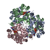
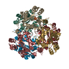
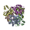
Deposited unit
A: Nucleoside diphosphate kinase
B: Nucleoside diphosphate kinase
C: Nucleoside diphosphate kinase
D: Nucleoside diphosphate kinase
E: Nucleoside diphosphate kinase
F: Nucleoside diphosphate kinase
G: Nucleoside diphosphate kinase
H: Nucleoside diphosphate kinase
I: Nucleoside diphosphate kinase
J: Nucleoside diphosphate kinase
K: Nucleoside diphosphate kinase
L: Nucleoside diphosphate kinase
M: Nucleoside diphosphate kinase
N: Nucleoside diphosphate kinase
Q: Nucleoside diphosphate kinase
R: Nucleoside diphosphate kinase
U: Nucleoside diphosphate kinase
V: Nucleoside diphosphate kinase
W: Nucleoside diphosphate kinase
X: Nucleoside diphosphate kinase
O: Nucleoside diphosphate kinase
P: Nucleoside diphosphate kinase
S: Nucleoside diphosphate kinase
T: Nucleoside diphosphate kinase Summary Component details
Theoretical mass Number of molelcules Total (without water) 412,480 24 Polymers 412,480 24 Non-polymers 0 0 Water 0 0
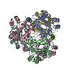
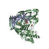
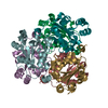
1
A: Nucleoside diphosphate kinase
B: Nucleoside diphosphate kinase
C: Nucleoside diphosphate kinase
D: Nucleoside diphosphate kinase
M: Nucleoside diphosphate kinase
N: Nucleoside diphosphate kinase Summary Component details Symmetry operations Calculated values
Theoretical mass Number of molelcules Total (without water) 103,120 6 Polymers 103,120 6 Non-polymers 0 0 Water 0
Type Name Symmetry operation Number identity operation 1_555 x,y,z 1
Buried area 16170 Å2 ΔGint -59 kcal/mol Surface area 33670 Å2 Method
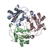
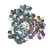
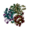
2
E: Nucleoside diphosphate kinase
F: Nucleoside diphosphate kinase
G: Nucleoside diphosphate kinase
H: Nucleoside diphosphate kinase
O: Nucleoside diphosphate kinase
P: Nucleoside diphosphate kinase Summary Component details Symmetry operations Calculated values
Theoretical mass Number of molelcules Total (without water) 103,120 6 Polymers 103,120 6 Non-polymers 0 0 Water 0
Type Name Symmetry operation Number identity operation 1_555 x,y,z 1
Buried area 16100 Å2 ΔGint -55 kcal/mol Surface area 33510 Å2 Method
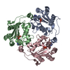
3
I: Nucleoside diphosphate kinase
J: Nucleoside diphosphate kinase
K: Nucleoside diphosphate kinase
L: Nucleoside diphosphate kinase
Q: Nucleoside diphosphate kinase
R: Nucleoside diphosphate kinase Summary Component details Symmetry operations Calculated values
Theoretical mass Number of molelcules Total (without water) 103,120 6 Polymers 103,120 6 Non-polymers 0 0 Water 0
Type Name Symmetry operation Number identity operation 1_555 x,y,z 1
Buried area 16050 Å2 ΔGint -59 kcal/mol Surface area 33820 Å2 Method
4
U: Nucleoside diphosphate kinase
V: Nucleoside diphosphate kinase
W: Nucleoside diphosphate kinase
X: Nucleoside diphosphate kinase
S: Nucleoside diphosphate kinase
T: Nucleoside diphosphate kinase Summary Component details Symmetry operations Calculated values
Theoretical mass Number of molelcules Total (without water) 103,120 6 Polymers 103,120 6 Non-polymers 0 0 Water 0
Type Name Symmetry operation Number identity operation 1_555 x,y,z 1
Buried area 16300 Å2 ΔGint -60 kcal/mol Surface area 33360 Å2 Method
Unit cell Length a, b, c (Å) 190.836, 169.470, 146.937 Angle α, β, γ (deg.) 90.00, 92.92, 90.00 Int Tables number 5 Space group name H-M C121
 Movie
Movie Controller

 Yorodumi
 Open data
 Basic information
 Components
 Keywords
 Function and homology information
 X-RAY DIFFRACTION /
  Authors
 China, 1items
  Citation
 Structure visualization
 Downloads & links
 Other downloads
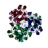

 PDBj
PDBj Assembly


 Sample preparation
 Processing