 Movie
Movie Controller

[English] 日本語
 Yorodumi
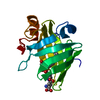
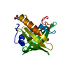
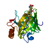
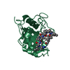
- PDB-5uwm: Matrix metalloproteinase-13 complexed with selective inhibitor co... -
+ Open data

- Basic information
| Entry | Database: PDB / ID: 5uwm | ||||||
|---|---|---|---|---|---|---|---|
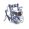
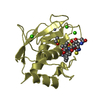
| Title | Matrix metalloproteinase-13 complexed with selective inhibitor compound (R)-17a | ||||||
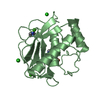
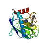
 Components Components | (Collagenase 3) x 2 | ||||||
 Keywords Keywords | hydrolase/hydrolase inhibitor / Metalloproteinase / collagenase / MMP-13 / hydrolase / hydrolase-hydrolase inhibitor complex | ||||||
| Function / homology |  Function and homology information Function and homology informationgrowth plate cartilage development / RUNX2 regulates genes involved in cell migration / endochondral ossification / Hydrolases; Acting on peptide bonds (peptidases); Metalloendopeptidases / bone morphogenesis / Assembly of collagen fibrils and other multimeric structures / bone mineralization / Activation of Matrix Metalloproteinases / response to amyloid-beta / Collagen degradation ...growth plate cartilage development / RUNX2 regulates genes involved in cell migration / endochondral ossification / Hydrolases; Acting on peptide bonds (peptidases); Metalloendopeptidases / bone morphogenesis / Assembly of collagen fibrils and other multimeric structures / bone mineralization / Activation of Matrix Metalloproteinases / response to amyloid-beta / Collagen degradation / collagen catabolic process / extracellular matrix disassembly / Degradation of the extracellular matrix / collagen binding / extracellular matrix organization / metalloendopeptidase activity / extracellular matrix / endopeptidase activity / serine-type endopeptidase activity / calcium ion binding / proteolysis / : / extracellular region / zinc ion binding Similarity search - Function | ||||||
| Biological species |  Homo sapiens (human) Homo sapiens (human) | ||||||
| Method |  X-RAY DIFFRACTION / SYNCHROTRON / MOLECULAR REPLACEMENT / Resolution: 1.62 Å X-RAY DIFFRACTION / SYNCHROTRON / MOLECULAR REPLACEMENT / Resolution: 1.62 Å | ||||||
 Authors Authors | Taylor, A.B. / Cao, X. / Hart, P.J. | ||||||
 Citation Citation | Journal: J. Med. Chem. / Year: 2017 Title: Structure-Based Design and Synthesis of Potent and Selective Matrix Metalloproteinase 13 Inhibitors. Authors: Choi, J.Y. / Fuerst, R. / Knapinska, A.M. / Taylor, A.B. / Smith, L. / Cao, X. / Hart, P.J. / Fields, G.B. / Roush, W.R. | ||||||
| History |
|
- Structure visualization
| Structure viewer | Molecule: MolmilJmol/JSmol |
|---|
- Downloads & links
-Download
| PDBx/mmCIF format | 5uwm.cif.gz | 158.4 KB | Display | PDBx/mmCIF format |
|---|---|---|---|---|
| PDB format | pdb5uwm.ent.gz | 123.1 KB | Display | PDB format |
| PDBx/mmJSON format | 5uwm.json.gz | Tree view | PDBx/mmJSON format | |
| Others |  Other downloads Other downloads |
-Validation report
| Arichive directory | https://data.pdbj.org/pub/pdb/validation_reports/uw/5uwmftp://data.pdbj.org/pub/pdb/validation_reports/uw/5uwm | HTTPS FTP |
|---|
-Related structure data
| Related structure data |  5uwkC  5uwlC  5uwnC  4l19S C: citing same article ( S: Starting model for refinement |
|---|---|
| Similar structure data |


-Links
 PDBj
PDBj


- Assembly
| Deposited unit | 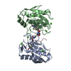
| ||||||||
|---|---|---|---|---|---|---|---|---|---|
| 1 | 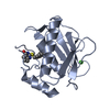
| ||||||||
| 2 | 
| ||||||||
| Unit cell |
|
-Components
-Protein / Protein/peptide , 2 types, 4 molecules ABCD
| #1: Protein | Mass: 19366.578 Da / Num. of mol.: 2 Source method: isolated from a genetically manipulated source Details: CATALYTIC DOMAIN (UNP RESIDUES 104-274) / Source: (gene. exp.) Homo sapiens (human) / Gene: MMP13 / Plasmid: PKA8H / Production host:  References: UniProt: P45452, Hydrolases; Acting on peptide bonds (peptidases); Metalloendopeptidases #2: Protein/peptide | Mass: 542.625 Da / Num. of mol.: 2 Source method: isolated from a genetically manipulated source Details: Proteolytic fragment / Source: (gene. exp.) Homo sapiens (human) / Plasmid: PKA8H / Production host: |
|---|
-Non-polymers , 4 types, 365 molecules 


| #3: Chemical | ChemComp-ZN /  Mass: 65.409 Da / Num. of mol.: 4 / Source method: obtained synthetically / Formula: Zn Mass: 65.409 Da / Num. of mol.: 4 / Source method: obtained synthetically / Formula: Zn#4: Chemical | ChemComp-CA /  Mass: 40.078 Da / Num. of mol.: 6 / Source method: obtained synthetically / Formula: Ca Mass: 40.078 Da / Num. of mol.: 6 / Source method: obtained synthetically / Formula: Ca#5: Chemical |  Mass: 480.579 Da / Num. of mol.: 2 / Source method: obtained synthetically / Formula: C25H28N4O4S Mass: 480.579 Da / Num. of mol.: 2 / Source method: obtained synthetically / Formula: C25H28N4O4S#6: Water | ChemComp-HOH / | Mass: 18.015 Da / Num. of mol.: 353 / Source method: isolated from a natural source / Formula: H2O |
|---|
-Experimental details
-Experiment
| Experiment | Method: X-RAY DIFFRACTION / Number of used crystals: 1 |
|---|
- Sample preparation
| Crystal | Density Matthews: 2.22 Å3/Da / Density % sol: 44.64 % |
|---|---|
| Crystal grow | Temperature: 295 K / Method: vapor diffusion, sitting drop / pH: 8.5 Details: 0.2 M lithium sulfate 0.1 M Tris:HCl pH 8.5, 30% polyethylene glycol 4000 |
-Data collection
| Diffraction | Mean temperature: 100 K |
|---|---|
| Diffraction source | Source: SYNCHROTRON / Site: APS  / Beamline: 24-ID-E / Wavelength: 0.97918 Å / Beamline: 24-ID-E / Wavelength: 0.97918 Å |
| Detector | Type: ADSC QUANTUM 315 / Detector: CCD / Date: Nov 14, 2015 |
| Radiation | Protocol: SINGLE WAVELENGTH / Monochromatic (M) / Laue (L): M / Scattering type: x-ray |
| Radiation wavelength | Wavelength: 0.97918 Å / Relative weight: 1 |
| Reflection | Resolution: 1.62→32.77 Å / Num. obs: 43442 / % possible obs: 99.7 % / Redundancy: 3.1 % / Biso Wilson estimate: 16.1 Å2 / Rpim(I) all: 0.046 / Rsym value: 0.069 / Net I/σ(I): 13.2 |
| Reflection shell | Resolution: 1.62→1.71 Å / Redundancy: 3.1 % / Mean I/σ(I) obs: 2 / Num. unique obs: 6358 / Rpim(I) all: 0.43 / Rsym value: 0.646 / % possible all: 99.9 |
- Processing
| Software |
| |||||||||||||||||||||||||||||||||||||||||||||||||||||||||||||||||||||||||||||||||||||||||||||||||||||||||
|---|---|---|---|---|---|---|---|---|---|---|---|---|---|---|---|---|---|---|---|---|---|---|---|---|---|---|---|---|---|---|---|---|---|---|---|---|---|---|---|---|---|---|---|---|---|---|---|---|---|---|---|---|---|---|---|---|---|---|---|---|---|---|---|---|---|---|---|---|---|---|---|---|---|---|---|---|---|---|---|---|---|---|---|---|---|---|---|---|---|---|---|---|---|---|---|---|---|---|---|---|---|---|---|---|---|---|
| Refinement | Method to determine structure: MOLECULAR REPLACEMENT Starting model: 4L19 Resolution: 1.62→32.77 Å / SU ML: 0.15 / Cross valid method: THROUGHOUT / σ(F): 1.97 / Phase error: 18.4
| |||||||||||||||||||||||||||||||||||||||||||||||||||||||||||||||||||||||||||||||||||||||||||||||||||||||||
| Solvent computation | Shrinkage radii: 0.9 Å / VDW probe radii: 1.11 Å | |||||||||||||||||||||||||||||||||||||||||||||||||||||||||||||||||||||||||||||||||||||||||||||||||||||||||
| Refinement step | Cycle: LAST / Resolution: 1.62→32.77 Å
| |||||||||||||||||||||||||||||||||||||||||||||||||||||||||||||||||||||||||||||||||||||||||||||||||||||||||
| Refine LS restraints |
| |||||||||||||||||||||||||||||||||||||||||||||||||||||||||||||||||||||||||||||||||||||||||||||||||||||||||
| LS refinement shell |
|
