[English] 日本語
Yorodumi
- PDB-3ts4: Human MMP12 in complex with L-glutamate motif inhibitor -

+
Open data


ID or keywords:

Loading...

-
Basic information

Entry
Database: PDB / ID: 3ts4
TitleHuman MMP12 in complex with L-glutamate motif inhibitor
ComponentsMacrophage metalloelastase
KeywordsHYDROLASE/HYDROLASE INHIBITOR / pseudo dipeptides / potent inhibitors / metzincin / Zinc protease / HYDROLASE-HYDROLASE INHIBITOR complex
Function / homology
Function and homology information


macrophage elastase / negative regulation of endothelial cell-matrix adhesion via fibronectin / bronchiole development / positive regulation of epithelial cell proliferation involved in wound healing / elastin catabolic process / regulation of defense response to virus by host / positive regulation of type I interferon-mediated signaling pathway / wound healing, spreading of epidermal cells / negative regulation of type I interferon-mediated signaling pathway / lung alveolus development ...macrophage elastase / negative regulation of endothelial cell-matrix adhesion via fibronectin / bronchiole development / positive regulation of epithelial cell proliferation involved in wound healing / elastin catabolic process / regulation of defense response to virus by host / positive regulation of type I interferon-mediated signaling pathway / wound healing, spreading of epidermal cells / negative regulation of type I interferon-mediated signaling pathway / lung alveolus development / positive regulation of interferon-alpha production / response to amyloid-beta / Collagen degradation / collagen catabolic process / extracellular matrix disassembly / core promoter sequence-specific DNA binding / collagen binding / extracellular matrix / Degradation of the extracellular matrix / extracellular matrix organization / metalloendopeptidase activity / cellular response to virus / protein import into nucleus / endopeptidase activity / sequence-specific DNA binding / serine-type endopeptidase activity / calcium ion binding / negative regulation of transcription by RNA polymerase II / positive regulation of transcription by RNA polymerase II / proteolysis / extracellular space / zinc ion binding / extracellular region / nucleus / cytoplasm
Similarity search - Function
Peptidoglycan binding-like / Hemopexin, conserved site / Hemopexin domain signature. / Hemopexin-like domain / Peptidase M10A, cysteine switch, zinc binding site / Matrixins cysteine switch. / Putative peptidoglycan binding domain / Hemopexin-like repeats / Hemopexin-like domain superfamily / Hemopexin ...Peptidoglycan binding-like / Hemopexin, conserved site / Hemopexin domain signature. / Hemopexin-like domain / Peptidase M10A, cysteine switch, zinc binding site / Matrixins cysteine switch. / Putative peptidoglycan binding domain / Hemopexin-like repeats / Hemopexin-like domain superfamily / Hemopexin / Hemopexin repeat profile. / Hemopexin-like repeats. / Peptidase M10A / Peptidase M10A, catalytic domain / Peptidase M10, metallopeptidase / Matrixin / PGBD-like superfamily / Peptidase, metallopeptidase / Zinc-dependent metalloprotease / Collagenase (Catalytic Domain) / Collagenase (Catalytic Domain) / Metallopeptidase, catalytic domain superfamily / Neutral zinc metallopeptidases, zinc-binding region signature. / 3-Layer(aba) Sandwich / Alpha Beta
Similarity search - Domain/homology
N-{3-[4-(4-PHENYLTHIOPHEN-2-YL)PHENYL]PROPANOYL}-L-ALPHA-GLUTAMYL-L-ALPHA-GLUTAMYL-AMIDE / Chem-EEG / IMIDAZOLE / Macrophage metalloelastase
Similarity search - Component
Biological speciesHomo sapiens (human)
MethodX-RAY DIFFRACTION / SYNCHROTRON / MOLECULAR REPLACEMENT / Resolution: 1.587 Å
AuthorsStura, E.A. / Dive, V. / Devel, L. / Czarny, B. / Beau, F. / Vera, L.
CitationJournal: J.Biol.Chem. / Year: 2012
Title: Simple pseudo-dipeptides with a P2' glutamate: a novel inhibitor family of matrix metalloproteases and other metzincins.
Authors: Devel, L. / Beau, F. / Amoura, M. / Vera, L. / Cassar-Lajeunesse, E. / Garcia, S. / Czarny, B. / Stura, E.A. / Dive, V.
History
DepositionSep 12, 2011Deposition site: RCSB / Processing site: RCSB
Revision 1.0Jun 20, 2012Provider: repository / Type: Initial release
Revision 1.1Oct 17, 2012Group: Database references
Revision 1.2Dec 12, 2012Group: Other
Revision 1.3Sep 13, 2023Group: Data collection / Database references ...Data collection / Database references / Derived calculations / Refinement description
Category: chem_comp_atom / chem_comp_bond ...chem_comp_atom / chem_comp_bond / database_2 / pdbx_initial_refinement_model / pdbx_struct_conn_angle / struct_conn / struct_ref_seq_dif / struct_site
Item: _database_2.pdbx_DOI / _database_2.pdbx_database_accession ..._database_2.pdbx_DOI / _database_2.pdbx_database_accession / _pdbx_struct_conn_angle.ptnr1_auth_comp_id / _pdbx_struct_conn_angle.ptnr1_auth_seq_id / _pdbx_struct_conn_angle.ptnr1_label_asym_id / _pdbx_struct_conn_angle.ptnr1_label_atom_id / _pdbx_struct_conn_angle.ptnr1_label_comp_id / _pdbx_struct_conn_angle.ptnr1_label_seq_id / _pdbx_struct_conn_angle.ptnr2_auth_comp_id / _pdbx_struct_conn_angle.ptnr2_auth_seq_id / _pdbx_struct_conn_angle.ptnr2_label_asym_id / _pdbx_struct_conn_angle.ptnr2_label_atom_id / _pdbx_struct_conn_angle.ptnr2_label_comp_id / _pdbx_struct_conn_angle.ptnr3_auth_comp_id / _pdbx_struct_conn_angle.ptnr3_auth_seq_id / _pdbx_struct_conn_angle.ptnr3_label_asym_id / _pdbx_struct_conn_angle.ptnr3_label_atom_id / _pdbx_struct_conn_angle.ptnr3_label_comp_id / _pdbx_struct_conn_angle.ptnr3_label_seq_id / _pdbx_struct_conn_angle.value / _struct_conn.pdbx_dist_value / _struct_conn.ptnr1_auth_comp_id / _struct_conn.ptnr1_auth_seq_id / _struct_conn.ptnr1_label_asym_id / _struct_conn.ptnr1_label_atom_id / _struct_conn.ptnr1_label_comp_id / _struct_conn.ptnr1_label_seq_id / _struct_conn.ptnr2_auth_comp_id / _struct_conn.ptnr2_auth_seq_id / _struct_conn.ptnr2_label_asym_id / _struct_conn.ptnr2_label_atom_id / _struct_conn.ptnr2_label_comp_id / _struct_ref_seq_dif.details / _struct_site.pdbx_auth_asym_id / _struct_site.pdbx_auth_comp_id / _struct_site.pdbx_auth_seq_id

-
Structure visualization

Structure viewerMolecule:
MolmilJmol/JSmol

Downloads & links

-
Assembly

Deposited unit
A: Macrophage metalloelastase
hetero molecules


Theoretical massNumber of molelcules
Total (without water)18,5949
Polymers17,6161
Non-polymers9788
Water3,837213
1


  • Idetical with deposited unit
  • defined by author&software
TypeNameSymmetry operationNumber
identity operation1_555x,y,z1
MethodPISA
Unit cell
Length a, b, c (Å)70.090, 63.650, 37.370
Angle α, β, γ (deg.)90.00, 90.00, 90.00
Int Tables number18
Space group name H-MP21212

-
Components

-
Protein , 1 types, 1 molecules A

#1: Protein Macrophage metalloelastase / MME / Macrophage elastase / ME / hME / Matrix metalloproteinase-12 / MMP-12


Mass: 17615.670 Da / Num. of mol.: 1 / Fragment: catalitic subunit (UNP RESIDUES 106-263) / Mutation: F171D
Source method: isolated from a genetically manipulated source
Source: (gene. exp.) Homo sapiens (human) / Gene: HME, MMP12 / Production host: Escherichia coli (E. coli) / Strain (production host): BL21 / References: UniProt: P39900, macrophage elastase

-
Non-polymers , 6 types, 221 molecules

#2: Chemical ChemComp-ZN / ZINC ION


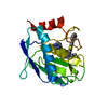
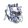
Mass: 65.409 Da / Num. of mol.: 2 / Source method: obtained synthetically / Formula: Zn
#3: Chemical ChemComp-CA / CALCIUM ION


Mass: 40.078 Da / Num. of mol.: 3 / Source method: obtained synthetically / Formula: Ca
#4: Chemical ChemComp-EEG / N-{3-[4-(4-phenylthiophen-2-yl)phenyl]propanoyl}-L-alpha-glutamyl-L-alpha-glutamyl-amide


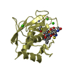
Type: Peptide-like / Class: Inhibitor / Mass: 565.637 Da / Num. of mol.: 1 / Source method: obtained synthetically / Formula: C29H31N3O7S
References: N-{3-[4-(4-PHENYLTHIOPHEN-2-YL)PHENYL]PROPANOYL}-L-ALPHA-GLUTAMYL-L-ALPHA-GLUTAMYL-AMIDE
#5: Chemical ChemComp-IMD / IMIDAZOLE / Imidazole


Mass: 69.085 Da / Num. of mol.: 1 / Source method: obtained synthetically / Formula: C3H5N2
#6: Chemical ChemComp-GOL / GLYCEROL / GLYCERIN / PROPANE-1,2,3-TRIOL / Glycerol


Mass: 92.094 Da / Num. of mol.: 1 / Source method: obtained synthetically / Formula: C3H8O3
#7: Water ChemComp-HOH / water / Water


Mass: 18.015 Da / Num. of mol.: 213 / Source method: isolated from a natural source / Formula: H2O

-
Experimental details

-
Experiment

ExperimentMethod: X-RAY DIFFRACTION / Number of used crystals: 1

-
Sample preparation

CrystalDensity Matthews: 2.37 Å3/Da / Density % sol: 48.01 %
Crystal growTemperature: 293 K / Method: vapor diffusion, sitting drop / pH: 9
Details: MMP12 AT 0.4 mM co-crystallization, with 100mM AHA and non-chelating inhibitor EEG at 1.1 mM with reservoir: 27% PEG 10K, 0.1M glycine. AHA removed during cryo-soak for 5 min in 30% MPEG2K, ...Details: MMP12 AT 0.4 mM co-crystallization, with 100mM AHA and non-chelating inhibitor EEG at 1.1 mM with reservoir: 27% PEG 10K, 0.1M glycine. AHA removed during cryo-soak for 5 min in 30% MPEG2K, 10% ethylene glycol, 200 mM imidazole malate, pH 8.5, VAPOR DIFFUSION, SITTING DROP, temperature 293K

-
Data collection

DiffractionMean temperature: 100 K
Diffraction sourceSource: SYNCHROTRON / Site: ESRF / Beamline: ID23-1 / Wavelength: 0.9919 Å
DetectorType: ADSC QUANTUM 315r / Detector: CCD / Date: Jul 20, 2009 / Details: bent cylindrical mirror
RadiationMonochromator: horizontally diffracting Si (111) monochromator and Pt coated mirrors in Kirkpatrick-Baez geometry for focusing
Protocol: SINGLE WAVELENGTH / Monochromatic (M) / Laue (L): M / Scattering type: x-ray
Radiation wavelengthWavelength: 0.9919 Å / Relative weight: 1
ReflectionResolution: 1.59→33 Å / Num. all: 23301 / Num. obs: 23208 / % possible obs: 99.6 % / Observed criterion σ(F): 0 / Observed criterion σ(I): 0 / Redundancy: 7.67 % / Biso Wilson estimate: 25.7 Å2 / Rmerge(I) obs: 0.099 / Rsym value: 0.081 / Net I/σ(I): 16.2
Reflection shell

Diffraction-ID: 1

Resolution (Å)Redundancy (%)Rmerge(I) obsMean I/σ(I) obsNum. unique allRsym value% possible all
1.59-1.687.580.6322.8936870.80598.8
4.72-336.860.01940.169930.04194.7
3.35-4.727.310.0240.1616510.04599.8
2.74-3.357.520.0332.3420710.058100
2.38-2.747.750.04824.4124350.078100
2.13-2.387.790.06818.6827400.108100
1.94-2.137.860.10213.429970.156100
1.8-1.947.840.2038.1332460.27100
1.68-1.87.780.3664.8734890.48299.9

-
Processing

Software
NameVersionClassification
DNAdata collection
MOLREPphasing
PHENIX(phenix.refine)refinement
XDSdata reduction
XSCALEdata scaling
RefinementMethod to determine structure: MOLECULAR REPLACEMENT
Starting model: 3LIK
Resolution: 1.587→32.976 Å / SU ML: 0.39 / Isotropic thermal model: anisotropic (TLS) / Cross valid method: THROUGHOUT / σ(F): 2.01 / Phase error: 19.06 / Stereochemistry target values: ML
RfactorNum. reflection% reflectionSelection details
Rfree0.203 1160 5 %RANDOM
Rwork0.1784 ---
obs0.1797 23181 99.51 %-
all-23295 --
Solvent computationShrinkage radii: 0.72 Å / VDW probe radii: 1 Å / Solvent model: FLAT BULK SOLVENT MODEL / Bsol: 53.451 Å2 / ksol: 0.333 e/Å3
Displacement parameters
Baniso -1Baniso -2Baniso -3
1-0.3654 Å2-0 Å2-0 Å2
2---0.1928 Å20 Å2
3----0.1726 Å2
Refinement stepCycle: LAST / Resolution: 1.587→32.976 Å
ProteinNucleic acidLigandSolventTotal
Num. atoms1246 0 56 213 1515
Refine LS restraints
Refine-IDTypeDev idealNumber
X-RAY DIFFRACTIONf_bond_d0.0161398
X-RAY DIFFRACTIONf_angle_d1.6121899
X-RAY DIFFRACTIONf_dihedral_angle_d18.681482
X-RAY DIFFRACTIONf_chiral_restr0.095188
X-RAY DIFFRACTIONf_plane_restr0.005253
LS refinement shell
Resolution (Å)Rfactor RfreeNum. reflection RfreeRfactor RworkNum. reflection RworkRefine-ID% reflection obs (%)
1.5871-1.65940.29661400.27622654X-RAY DIFFRACTION98
1.6594-1.74680.25711440.22712730X-RAY DIFFRACTION100
1.7468-1.85630.2061430.19842736X-RAY DIFFRACTION100
1.8563-1.99960.18571430.17762724X-RAY DIFFRACTION100
1.9996-2.20080.2141460.1772755X-RAY DIFFRACTION100
2.2008-2.51910.21041440.18132742X-RAY DIFFRACTION100
2.5191-3.17340.22221480.17572806X-RAY DIFFRACTION100
3.1734-32.98280.17391520.1592874X-RAY DIFFRACTION98
Refinement TLS params.Method: refined / Origin x: 7.2644 Å / Origin y: 15.892 Å / Origin z: 3.0981 Å
111213212223313233
T0.0757 Å20.0174 Å20.0131 Å2-0.0471 Å20.0461 Å2--0.0157 Å2
L6.4628 °20.174 °20.6073 °2-1.5624 °20.004 °2--1.1953 °2
S-0.0246 Å °-0.3765 Å °-0.3007 Å °0.077 Å °0.0391 Å °0.0268 Å °0.0388 Å °-0.0831 Å °-0.0129 Å °
Refinement TLS groupSelection details: all

+
About Yorodumi

-
News

-
Feb 9, 2022. New format data for meta-information of EMDB entries

New format data for meta-information of EMDB entries

  • Version 3 of the EMDB header file is now the official format.
  • The previous official version 1.9 will be removed from the archive.

Related info.:EMDB header

External links:wwPDB to switch to version 3 of the EMDB data model

-
Aug 12, 2020. Covid-19 info

Covid-19 info

URL: https://pdbj.org/emnavi/covid19.php

New page: Covid-19 featured information page in EM Navigator.

Related info.:Covid-19 info / Mar 5, 2020. Novel coronavirus structure data

+
Mar 5, 2020. Novel coronavirus structure data

Novel coronavirus structure data

Related info.:Yorodumi Speices / Aug 12, 2020. Covid-19 info

External links:COVID-19 featured content - PDBj / Molecule of the Month (242):Coronavirus Proteases

+
Jan 31, 2019. EMDB accession codes are about to change! (news from PDBe EMDB page)

EMDB accession codes are about to change! (news from PDBe EMDB page)

  • The allocation of 4 digits for EMDB accession codes will soon come to an end. Whilst these codes will remain in use, new EMDB accession codes will include an additional digit and will expand incrementally as the available range of codes is exhausted. The current 4-digit format prefixed with “EMD-” (i.e. EMD-XXXX) will advance to a 5-digit format (i.e. EMD-XXXXX), and so on. It is currently estimated that the 4-digit codes will be depleted around Spring 2019, at which point the 5-digit format will come into force.
  • The EM Navigator/Yorodumi systems omit the EMD- prefix.

Related info.:Q: What is EMD? / ID/Accession-code notation in Yorodumi/EM Navigator

External links:EMDB Accession Codes are Changing Soon! / Contact to PDBj

+
Jul 12, 2017. Major update of PDB

Major update of PDB

  • wwPDB released updated PDB data conforming to the new PDBx/mmCIF dictionary.
  • This is a major update changing the version number from 4 to 5, and with Remediation, in which all the entries are updated.
  • In this update, many items about electron microscopy experimental information are reorganized (e.g. em_software).
  • Now, EM Navigator and Yorodumi are based on the updated data.

External links:wwPDB Remediation / Enriched Model Files Conforming to OneDep Data Standards Now Available in the PDB FTP Archive

-
Yorodumi

Thousand views of thousand structures

  • Yorodumi is a browser for structure data from EMDB, PDB, SASBDB, etc.
  • This page is also the successor to EM Navigator detail page, and also detail information page/front-end page for Omokage search.
  • The word "yorodu" (or yorozu) is an old Japanese word meaning "ten thousand". "mi" (miru) is to see.

Related info.:EMDB / PDB / SASBDB / Comparison of 3 databanks / Yorodumi Search / Aug 31, 2016. New EM Navigator & Yorodumi / Yorodumi Papers / Jmol/JSmol / Function and homology information / Changes in new EM Navigator and Yorodumi

Read more