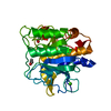
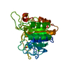
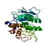
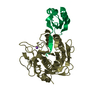
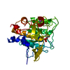
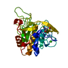
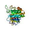
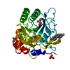
 Movie
Movie Controller


+ Open data

- Basic information
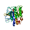
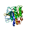
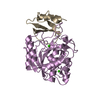
| Entry | Database: PDB / ID: 1gnv | ||||||
|---|---|---|---|---|---|---|---|
| Title | CALCIUM INDEPENDENT SUBTILISIN BPN' MUTANT | ||||||
 Components Components | SUBTILISIN BPN' | ||||||
 Keywords Keywords | HYDROLASE / SERINE PROTEINASE | ||||||
| Function / homology |  Function and homology information Function and homology informationsubtilisin / sporulation resulting in formation of a cellular spore / fibrinolysis / serine-type endopeptidase activity / proteolysis / extracellular region / metal ion binding Similarity search - Function | ||||||
| Biological species |  | ||||||
| Method |  X-RAY DIFFRACTION / MOLECULAR REPLACEMENT / Resolution: 1.9 Å X-RAY DIFFRACTION / MOLECULAR REPLACEMENT / Resolution: 1.9 Å | ||||||
 Authors Authors | Almog, O. / Gilliland, G.L. | ||||||
 Citation Citation | Journal: J.Biol.Chem. / Year: 2002 Title: Structural Basis of Thermostability. Analysis of Stabilizing Mutations in Subtilisin Bpn'. Authors: Almog, O. / Gallagher, D.T. / Ladner, J.E. / Strausberg, S. / Alexander, P. / Bryan, P. / Gilliland, G.L. | ||||||
| History |
|
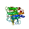
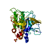
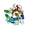
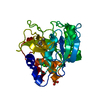
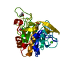
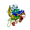
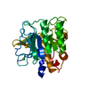
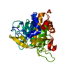
- Structure visualization
| Structure viewer | Molecule: MolmilJmol/JSmol |
|---|
- Downloads & links
-Download
| PDBx/mmCIF format | 1gnv.cif.gz | 64.5 KB | Display | PDBx/mmCIF format |
|---|---|---|---|---|
| PDB format | pdb1gnv.ent.gz | 45.6 KB | Display | PDB format |
| PDBx/mmJSON format | 1gnv.json.gz | Tree view | PDBx/mmJSON format | |
| Others |  Other downloads Other downloads |
-Validation report
| Arichive directory | https://data.pdbj.org/pub/pdb/validation_reports/gn/1gnvftp://data.pdbj.org/pub/pdb/validation_reports/gn/1gnv | HTTPS FTP |
|---|
-Related structure data
| Related structure data | 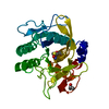 1gnsC  1suaS C: citing same article ( S: Starting model for refinement |
|---|---|
| Similar structure data |


-Links
 PDBj
PDBj

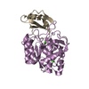
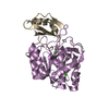
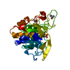
- Assembly
| Deposited unit | 
| ||||||||
|---|---|---|---|---|---|---|---|---|---|
| 1 |
| ||||||||
| Unit cell |
|
-Components
| #1: Protein | Mass: 26771.629 Da / Num. of mol.: 1 / Mutation: YES Source method: isolated from a genetically manipulated source Source: (gene. exp.) |
|---|---|
| #2: Water | ChemComp-HOH /  Mass: 18.015 Da / Num. of mol.: 133 / Source method: isolated from a natural source / Formula: H2O Mass: 18.015 Da / Num. of mol.: 133 / Source method: isolated from a natural source / Formula: H2O |
| Compound details | DELETION, RESIDUES 176-184. OTHER MUTATIONS: Q103K, S104C, P106S, K144N, M151F, A174L, Q174C, ...DELETION, RESIDUES 176-184. OTHER MUTATIONS: Q103K, S104C, P106S, K144N, M151F, A174L, Q174C, Y318K, N319S, Q372E |
-Experimental details
-Experiment
| Experiment | Method: X-RAY DIFFRACTION / Number of used crystals: 1 |
|---|
- Sample preparation
| Crystal | Density Matthews: 2.53 Å3/Da / Density % sol: 51.35 % | ||||||||||||||||||||||||||||||
|---|---|---|---|---|---|---|---|---|---|---|---|---|---|---|---|---|---|---|---|---|---|---|---|---|---|---|---|---|---|---|---|
| Crystal grow | pH: 7.5 / Details: 23% PEG 4K, O.2M AMMONIUM SULFATE, pH 7.50 | ||||||||||||||||||||||||||||||
| Crystal grow | *PLUS Temperature: 20 ℃ / pH: 9 / Method: vapor diffusion, hanging drop | ||||||||||||||||||||||||||||||
| Components of the solutions | *PLUS
|
-Data collection
| Diffraction | Mean temperature: 293 K |
|---|---|
| Diffraction source | Source: ROTATING ANODE / Type: RIGAKU RU200 / Wavelength: 1.5418 |
| Detector | Type: SIEMENS MULTIWIRE / Detector: AREA DETECTOR / Date: Jul 15, 1994 |
| Radiation | Monochromator: NI/FILTER / Protocol: SINGLE WAVELENGTH / Monochromatic (M) / Laue (L): M / Scattering type: x-ray |
| Radiation wavelength | Wavelength: 1.5418 Å / Relative weight: 1 |
| Reflection | Resolution: 1.9→8 Å / Num. obs: 17076 / % possible obs: 86 % / Observed criterion σ(I): 2 / Redundancy: 3 % / Rmerge(I) obs: 0.088 / Net I/σ(I): 6 |
| Reflection shell | Rmerge(I) obs: 0.088 |
| Reflection | *PLUS Num. all: 19754 / Num. obs: 17067 / Num. measured all: 72438 |
- Processing
| Software |
| |||||||||||||||||||||||||||||||||||||||||||||||||||||||||||||||
|---|---|---|---|---|---|---|---|---|---|---|---|---|---|---|---|---|---|---|---|---|---|---|---|---|---|---|---|---|---|---|---|---|---|---|---|---|---|---|---|---|---|---|---|---|---|---|---|---|---|---|---|---|---|---|---|---|---|---|---|---|---|---|---|---|
| Refinement | Method to determine structure: MOLECULAR REPLACEMENT Starting model: PDB ENTRY 1SUA Resolution: 1.9→8 Å / σ(F): 2
| |||||||||||||||||||||||||||||||||||||||||||||||||||||||||||||||
| Refinement step | Cycle: LAST / Resolution: 1.9→8 Å
| |||||||||||||||||||||||||||||||||||||||||||||||||||||||||||||||
| Refine LS restraints |
| |||||||||||||||||||||||||||||||||||||||||||||||||||||||||||||||
| Refinement | *PLUS Rfactor all: 0.176 | |||||||||||||||||||||||||||||||||||||||||||||||||||||||||||||||
| Solvent computation | *PLUS | |||||||||||||||||||||||||||||||||||||||||||||||||||||||||||||||
| Displacement parameters | *PLUS | |||||||||||||||||||||||||||||||||||||||||||||||||||||||||||||||
| Refine LS restraints | *PLUS Type: p_bond_d / Dev ideal: 0.02 |
