 Movie
Movie Controller

[English] 日本語
 Yorodumi
- PDB-2sni: STRUCTURAL COMPARISON OF TWO SERINE PROTEINASE-PROTEIN INHIBITOR ... -
+ Open data

- Basic information
| Entry | Database: PDB / ID: 2sni | |||||||||
|---|---|---|---|---|---|---|---|---|---|---|
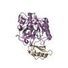
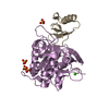
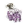
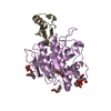
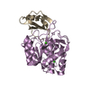
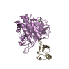
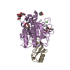
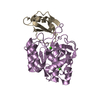
| Title | STRUCTURAL COMPARISON OF TWO SERINE PROTEINASE-PROTEIN INHIBITOR COMPLEXES. EGLIN-C-SUBTILISIN CARLSBERG AND CI-2-SUBTILISIN NOVO | |||||||||
 Components Components |
| |||||||||
 Keywords Keywords | COMPLEX (PROTEINASE/INHIBITOR) / COMPLEX (PROTEINASE-INHIBITOR) / COMPLEX (PROTEINASE-INHIBITOR) complex | |||||||||
| Function / homology |  Function and homology information Function and homology informationsubtilisin / sporulation resulting in formation of a cellular spore / fibrinolysis / serine-type endopeptidase inhibitor activity / response to wounding / serine-type endopeptidase activity / proteolysis / extracellular region / metal ion binding Similarity search - Function | |||||||||
| Biological species |   | |||||||||
| Method |  X-RAY DIFFRACTION / Resolution: 2.1 Å X-RAY DIFFRACTION / Resolution: 2.1 Å | |||||||||
 Authors Authors | Mcphalen, C.A. / James, M.N.G. | |||||||||
 Citation Citation | Journal: Biochemistry / Year: 1988 Title: Structural comparison of two serine proteinase-protein inhibitor complexes: eglin-c-subtilisin Carlsberg and CI-2-subtilisin Novo. Authors: McPhalen, C.A. / James, M.N. #1: Journal: Biochemistry / Year: 1987Title: Crystal and Molecular Structure of the Serine Proteinase Inhibitor Ci-2 from Barley Seeds Authors: Mcphalen, C.A. / James, M.N.G. #2: Journal: Proc.Natl.Acad.Sci.USA / Year: 1985Title: Crystal and Molecular Structure of Chymotrypsin Inhibitor 2 from Barley Seeds in Complex with Subtilisin Novo Authors: Mcphalen, C.A. / Svendsen, I. / Jonassen, I. / James, M.N.G. | |||||||||
| History |
| |||||||||
| Remark 700 | SHEET THE CROSS-OVER CONNECTION BETWEEN STRANDS 1 AND 2 OF SHEET S1E IS LEFT-HANDED. THE BETA-SHEET ...SHEET THE CROSS-OVER CONNECTION BETWEEN STRANDS 1 AND 2 OF SHEET S1E IS LEFT-HANDED. THE BETA-SHEET OF THE INHIBITOR IS IRREGULAR , WITH WELL-ORDERED WATER MOLECULES PROVIDING ALL BUT ONE HYDROGEN-BONDING BRIDGE BETWEEN STRANDS 2 AND 3. SEE THE PAPER CITED ON THE *JRNL* RECORDS ABOVE. |
- Structure visualization
| Structure viewer | Molecule: MolmilJmol/JSmol |
|---|
- Downloads & links
-Download
| PDBx/mmCIF format | 2sni.cif.gz | 78.2 KB | Display | PDBx/mmCIF format |
|---|---|---|---|---|
| PDB format | pdb2sni.ent.gz | 57.5 KB | Display | PDB format |
| PDBx/mmJSON format | 2sni.json.gz | Tree view | PDBx/mmJSON format | |
| Others |  Other downloads Other downloads |
-Validation report
| Arichive directory | https://data.pdbj.org/pub/pdb/validation_reports/sn/2sniftp://data.pdbj.org/pub/pdb/validation_reports/sn/2sni | HTTPS FTP |
|---|
-Related structure data


-Links
 PDBj
PDBj

- Assembly
| Deposited unit | 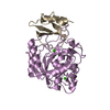
| ||||||||
|---|---|---|---|---|---|---|---|---|---|
| 1 |
| ||||||||
| Unit cell |
| ||||||||
| Atom site foot note | 1: SEE REMARK 7. / 2: RESIDUE CYS E 168 IS A CIS PROLINE. |
-Components
| #1: Protein | Mass: 27551.541 Da / Num. of mol.: 1 Source method: isolated from a genetically manipulated source Source: (gene. exp.) | ||||
|---|---|---|---|---|---|
| #2: Protein | Mass: 9264.659 Da / Num. of mol.: 1 Source method: isolated from a genetically manipulated source Source: (gene. exp.) | ||||
| #3: Chemical |   Mass: 40.078 Da / Num. of mol.: 2 / Source method: obtained synthetically / Formula: Ca Mass: 40.078 Da / Num. of mol.: 2 / Source method: obtained synthetically / Formula: Ca#4: Water | ChemComp-HOH / |  Mass: 18.015 Da / Num. of mol.: 168 / Source method: isolated from a natural source / Formula: H2O Mass: 18.015 Da / Num. of mol.: 168 / Source method: isolated from a natural source / Formula: H2OSequence details | THE ORDER OF THE FIRST FOUR RESIDUES OF CHAIN *I* IS UNKNOWN. | |
-Experimental details
-Experiment
| Experiment | Method: X-RAY DIFFRACTION |
|---|
- Sample preparation
| Crystal | Density Matthews: 2.17 Å3/Da / Density % sol: 43.35 % | ||||||||||||||||||
|---|---|---|---|---|---|---|---|---|---|---|---|---|---|---|---|---|---|---|---|
| Crystal grow | *PLUS pH: 5.5 / Method: vapor diffusion, hanging dropDetails: McPhalen, C.A., (1985) Proc. Natl. Acad. Sci. USA., 82, 7242. | ||||||||||||||||||
| Components of the solutions | *PLUS
|
- Processing
| Software | Name: PROLSQ / Classification: refinement | ||||||||||||||||||||||||||||||||||||||||||||||||||||||||||||||||||||||||||||||||||||
|---|---|---|---|---|---|---|---|---|---|---|---|---|---|---|---|---|---|---|---|---|---|---|---|---|---|---|---|---|---|---|---|---|---|---|---|---|---|---|---|---|---|---|---|---|---|---|---|---|---|---|---|---|---|---|---|---|---|---|---|---|---|---|---|---|---|---|---|---|---|---|---|---|---|---|---|---|---|---|---|---|---|---|---|---|---|
| Refinement | Resolution: 2.1→8 Å / σ(I): 1 Details: WATER MOLECULES WITH SEQUENCE NUMBERS 600 - 602 AND B VALUES OF 0.0 WERE ADDED AT THE POSITIONS OF STRONG PEAKS IN THE FINAL DIFFERENCE MAP, AND THEIR POSITIONS HAVE NOT BEEN REFINED.
| ||||||||||||||||||||||||||||||||||||||||||||||||||||||||||||||||||||||||||||||||||||
| Refinement step | Cycle: LAST / Resolution: 2.1→8 Å
| ||||||||||||||||||||||||||||||||||||||||||||||||||||||||||||||||||||||||||||||||||||
| Refine LS restraints |
|
