 Movie
Movie Controller

[English] 日本語
 Yorodumi
- PDB-1lw6: Crystal Structure of the Complex of Subtilisin BPN' with Chymotry... -
+ Open data

- Basic information
| Entry | Database: PDB / ID: 1lw6 | ||||||
|---|---|---|---|---|---|---|---|
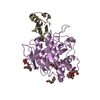
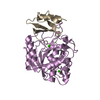
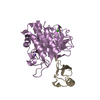
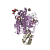
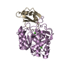
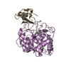
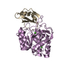
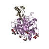
| Title | Crystal Structure of the Complex of Subtilisin BPN' with Chymotrypsin Inhibitor 2 at 1.5 Angstrom Resolution | ||||||
 Components Components |
| ||||||
 Keywords Keywords | HYDROLASE / SERINE PROTEASE / INHIBITOR | ||||||
| Function / homology |  Function and homology information Function and homology informationsubtilisin / sporulation resulting in formation of a cellular spore / fibrinolysis / serine-type endopeptidase inhibitor activity / response to wounding / serine-type endopeptidase activity / proteolysis / extracellular region / metal ion binding Similarity search - Function | ||||||
| Biological species |   | ||||||
| Method |  X-RAY DIFFRACTION / SYNCHROTRON / MOLECULAR REPLACEMENT / Resolution: 1.5 Å X-RAY DIFFRACTION / SYNCHROTRON / MOLECULAR REPLACEMENT / Resolution: 1.5 Å | ||||||
 Authors Authors | Radisky, E.S. / Koshland JR., D.E. | ||||||
 Citation Citation | Journal: Proc.Natl.Acad.Sci.USA / Year: 2002 Title: A clogged gutter mechanism for protease inhibitors. Authors: Radisky, E.S. / Koshland Jr., D.E. | ||||||
| History |
|
- Structure visualization
| Structure viewer | Molecule: MolmilJmol/JSmol |
|---|
- Downloads & links
-Download
| PDBx/mmCIF format | 1lw6.cif.gz | 89 KB | Display | PDBx/mmCIF format |
|---|---|---|---|---|
| PDB format | pdb1lw6.ent.gz | 65 KB | Display | PDB format |
| PDBx/mmJSON format | 1lw6.json.gz | Tree view | PDBx/mmJSON format | |
| Others |  Other downloads Other downloads |
-Validation report
| Arichive directory | https://data.pdbj.org/pub/pdb/validation_reports/lw/1lw6ftp://data.pdbj.org/pub/pdb/validation_reports/lw/1lw6 | HTTPS FTP |
|---|
-Related structure data
| Related structure data |  2sniS S: Starting model for refinement |
|---|---|
| Similar structure data |


-Links
 PDBj
PDBj

- Assembly
| Deposited unit | 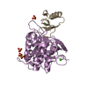
| ||||||||
|---|---|---|---|---|---|---|---|---|---|
| 1 |
| ||||||||
| Unit cell |
|
-Components
| #1: Protein | Mass: 28381.396 Da / Num. of mol.: 1 Source method: isolated from a genetically manipulated source Source: (gene. exp.) | ||
|---|---|---|---|
| #2: Protein | Mass: 7255.586 Da / Num. of mol.: 1 / Mutation: E45A Source method: isolated from a genetically manipulated source Source: (gene. exp.) | ||
| #3: Chemical | ChemComp-CA /   Mass: 40.078 Da / Num. of mol.: 1 / Source method: obtained synthetically / Formula: Ca Mass: 40.078 Da / Num. of mol.: 1 / Source method: obtained synthetically / Formula: Ca | ||
| #4: Chemical | ChemComp-SO4 /   Mass: 96.063 Da / Num. of mol.: 4 / Source method: obtained synthetically / Formula: SO4 Mass: 96.063 Da / Num. of mol.: 4 / Source method: obtained synthetically / Formula: SO4#5: Water | ChemComp-HOH / |  Mass: 18.015 Da / Num. of mol.: 505 / Source method: isolated from a natural source / Formula: H2O Mass: 18.015 Da / Num. of mol.: 505 / Source method: isolated from a natural source / Formula: H2O |
-Experimental details
-Experiment
| Experiment | Method: X-RAY DIFFRACTION / Number of used crystals: 1 |
|---|
- Sample preparation
| Crystal | Density Matthews: 2.56 Å3/Da / Density % sol: 51.91 % | ||||||||||||||||||||||||||||||||||||||||||
|---|---|---|---|---|---|---|---|---|---|---|---|---|---|---|---|---|---|---|---|---|---|---|---|---|---|---|---|---|---|---|---|---|---|---|---|---|---|---|---|---|---|---|---|
| Crystal grow | Temperature: 277 K / Method: vapor diffusion, hanging drop / pH: 6.5 Details: PEG 8000, ammonium sulfate, sodium cacodylate, pH 6.5, VAPOR DIFFUSION, HANGING DROP, temperature 277K | ||||||||||||||||||||||||||||||||||||||||||
| Crystal grow | *PLUS Temperature: 4 ℃ / pH: 5.8 | ||||||||||||||||||||||||||||||||||||||||||
| Components of the solutions | *PLUS
|
-Data collection
| Diffraction | Mean temperature: 100 K |
|---|---|
| Diffraction source | Source: SYNCHROTRON / Site: ALS  / Beamline: 5.0.1 / Wavelength: 1 Å / Beamline: 5.0.1 / Wavelength: 1 Å |
| Detector | Type: ADSC QUANTUM 4 / Detector: CCD / Date: Jul 21, 2001 |
| Radiation | Protocol: SINGLE WAVELENGTH / Monochromatic (M) / Laue (L): M / Scattering type: x-ray |
| Radiation wavelength | Wavelength: 1 Å / Relative weight: 1 |
| Reflection | Resolution: 1.5→29.66 Å / Num. all: 57529 / Num. obs: 57374 / % possible obs: 96.8 % / Observed criterion σ(F): -3 / Observed criterion σ(I): -3 / Redundancy: 6.5 % / Biso Wilson estimate: 12.4 Å2 / Rsym value: 0.072 / Net I/σ(I): 15.3 |
| Reflection shell | Resolution: 1.5→1.58 Å / Redundancy: 5 % / Mean I/σ(I) obs: 4.3 / Num. unique all: 7527 / Rsym value: 0.34 / % possible all: 88.9 |
| Reflection | *PLUS Highest resolution: 1.5 Å / Lowest resolution: 29.7 Å / Rmerge(I) obs: 0.072 |
| Reflection shell | *PLUS % possible obs: 88.9 % / Rmerge(I) obs: 0.34 |
- Processing
| Software |
| |||||||||||||||||||||||||
|---|---|---|---|---|---|---|---|---|---|---|---|---|---|---|---|---|---|---|---|---|---|---|---|---|---|---|
| Refinement | Method to determine structure: MOLECULAR REPLACEMENT Starting model: PDB ENTRY 2SNI Resolution: 1.5→29.66 Å / σ(F): -3 / Stereochemistry target values: Engh & Huber
| |||||||||||||||||||||||||
| Displacement parameters | Biso mean: 12.71 Å2 | |||||||||||||||||||||||||
| Refinement step | Cycle: LAST / Resolution: 1.5→29.66 Å
| |||||||||||||||||||||||||
| Refine LS restraints |
| |||||||||||||||||||||||||
| Refinement | *PLUS Highest resolution: 1.5 Å / Lowest resolution: 29.7 Å / Rfactor all: 0.17 / Rfactor Rfree: 0.188 / Rfactor Rwork: 0.169 | |||||||||||||||||||||||||
| Solvent computation | *PLUS | |||||||||||||||||||||||||
| Displacement parameters | *PLUS | |||||||||||||||||||||||||
| Refine LS restraints | *PLUS
|
