 Movie
Movie Controller

[English] 日本語
 Yorodumi
- PDB-1cse: THE HIGH-RESOLUTION X-RAY CRYSTAL STRUCTURE OF THE COMPLEX FORMED... -
+ Open data

- Basic information
| Entry | Database: PDB / ID: 1cse | ||||||
|---|---|---|---|---|---|---|---|
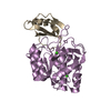
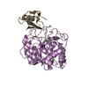
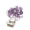
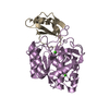
| Title | THE HIGH-RESOLUTION X-RAY CRYSTAL STRUCTURE OF THE COMPLEX FORMED BETWEEN SUBTILISIN CARLSBERG AND EGLIN C, AN ELASTASE INHIBITOR FROM THE LEECH HIRUDO MEDICINALIS. STRUCTURAL ANALYSIS, SUBTILISIN STRUCTURE AND INTERFACE GEOMETRY | ||||||
 Components Components |
| ||||||
 Keywords Keywords | COMPLEX(SERINE PROTEINASE-INHIBITOR) | ||||||
| Function / homology |  Function and homology information Function and homology informationsubtilisin / serine-type endopeptidase inhibitor activity / response to wounding / serine-type endopeptidase activity / proteolysis / : / metal ion binding Similarity search - Function | ||||||
| Biological species |   Hirudo medicinalis (medicinal leech) Hirudo medicinalis (medicinal leech) | ||||||
| Method |  X-RAY DIFFRACTION / Resolution: 1.2 Å X-RAY DIFFRACTION / Resolution: 1.2 Å | ||||||
 Authors Authors | Bode, W. | ||||||
 Citation Citation | Journal: Eur.J.Biochem. / Year: 1987 Title: The high-resolution X-ray crystal structure of the complex formed between subtilisin Carlsberg and eglin c, an elastase inhibitor from the leech Hirudo medicinalis. Structural analysis, ...Title: The high-resolution X-ray crystal structure of the complex formed between subtilisin Carlsberg and eglin c, an elastase inhibitor from the leech Hirudo medicinalis. Structural analysis, subtilisin structure and interface geometry. Authors: Bode, W. / Papamokos, E. / Musil, D. #1: Journal: Embo J. / Year: 1986Title: Refined 1.2 Angstroms Crystal Structure of the Complex Formed between Subtilisin Carlsberg and the Inhibitor Eglin C. Molecular Structure of Eglin and its Detailed Interaction with Subtilisin Authors: Bode, W. / Papamokos, E. / Musil, D. / Seemueller, U. / Fritz, H. #2: Journal: FEBS Lett. / Year: 1985Title: Crystal and Molecular Structure of the Inhibitor Eglin from Leeches in Complex with Subtilisin Carlsberg Authors: Mcphalen, C.A. / Schnebli, H.P. / James, M.N.G. | ||||||
| History |
|
- Structure visualization
| Structure viewer | Molecule: MolmilJmol/JSmol |
|---|
- Downloads & links
-Download
| PDBx/mmCIF format | 1cse.cif.gz | 85.5 KB | Display | PDBx/mmCIF format |
|---|---|---|---|---|
| PDB format | pdb1cse.ent.gz | 63.8 KB | Display | PDB format |
| PDBx/mmJSON format | 1cse.json.gz | Tree view | PDBx/mmJSON format | |
| Others |  Other downloads Other downloads |
-Validation report
| Arichive directory | https://data.pdbj.org/pub/pdb/validation_reports/cs/1cseftp://data.pdbj.org/pub/pdb/validation_reports/cs/1cse | HTTPS FTP |
|---|
-Related structure data
| Similar structure data |
|---|


-Links
 PDBj
PDBj

- Assembly
| Deposited unit | 
| ||||||||
|---|---|---|---|---|---|---|---|---|---|
| 1 |
| ||||||||
| Unit cell |
| ||||||||
| Atom site foot note | 1: RESIDUE PRO 168 IS A CIS PROLINE. / 2: RESIDUE THR 211 IS A CIS THREONINE. / 3: SEE REMARK 3. |
-Components
| #1: Protein | Mass: 27306.199 Da / Num. of mol.: 1 Source method: isolated from a genetically manipulated source Source: (gene. exp.) References: UniProt: P00780, UniProt: B0FXJ2*PLUS, subtilisin | ||
|---|---|---|---|
| #2: Protein | Mass: 8099.025 Da / Num. of mol.: 1 Source method: isolated from a genetically manipulated source Source: (gene. exp.) Hirudo medicinalis (medicinal leech) / References: UniProt: P01051 | ||
| #3: Chemical |   Mass: 40.078 Da / Num. of mol.: 2 / Source method: obtained synthetically / Formula: Ca Mass: 40.078 Da / Num. of mol.: 2 / Source method: obtained synthetically / Formula: Ca#4: Water | ChemComp-HOH / |  Mass: 18.015 Da / Num. of mol.: 432 / Source method: isolated from a natural source / Formula: H2O Mass: 18.015 Da / Num. of mol.: 432 / Source method: isolated from a natural source / Formula: H2O |
-Experimental details
-Experiment
| Experiment | Method: X-RAY DIFFRACTION |
|---|
- Sample preparation
| Crystal | Density Matthews: 2.29 Å3/Da / Density % sol: 46.4 % |
|---|---|
| Crystal grow | *PLUS Temperature: 20 ℃ / pH: 6.5 / Method: vapor diffusion, hanging drop / Details: seeding |
| Components of the solutions | *PLUS Conc.: 4-5 % / Common name: PEG |
-Data collection
| Reflection | *PLUS Highest resolution: 1.2 Å / Num. obs: 46280 / % possible obs: 45 % / Num. measured all: 94661 / Rmerge(I) obs: 0.092 |
|---|
- Processing
| Software | Name: EREF / Classification: refinement | ||||||||||||
|---|---|---|---|---|---|---|---|---|---|---|---|---|---|
| Refinement | Resolution: 1.2→10 Å Details: RESIDUE THR 211 IS A CIS THREONINE. IT WAS ORIGINALLY MODELLED AS A TRANS CONFORMER. AS OUTLINED IN REFERENCE 1 ABOVE, HOWEVER, THE MAIN CHAIN ANGLES OF THIS RESIDUE WERE OUTSIDE THE ALLOWED ...Details: RESIDUE THR 211 IS A CIS THREONINE. IT WAS ORIGINALLY MODELLED AS A TRANS CONFORMER. AS OUTLINED IN REFERENCE 1 ABOVE, HOWEVER, THE MAIN CHAIN ANGLES OF THIS RESIDUE WERE OUTSIDE THE ALLOWED REGIONS AND THE FIT TO THE ELECTRON DENSITY WAS STILL INSUFFICIENT. THE CIS CONFORMER GIVEN IN THIS ENTRY FITS MUCH BETTER. THE MODIFIED MODEL HAS BEEN SUBJECTED TO TWO FURTHER MINICYCLES OF POSITIONAL AND B FACTOR REFINEMENT WITHOUT GROSS CONFORMATION CHANGES AND WITHOUT AFFECTING THE R VALUE. TWO SITES PROBABLY OCCUPIED BY CALCIUM IONS AND 432 SOLVENT MOLECULES WERE LOCATED. FOR THESE 434 NON-PROTEIN ATOMS REFINED INDIVIDUAL OCCUPANCIES ARE GIVEN.
| ||||||||||||
| Refinement step | Cycle: LAST / Resolution: 1.2→10 Å
| ||||||||||||
| Refine LS restraints |
| ||||||||||||
| Software | *PLUS Name: EREF / Classification: refinement | ||||||||||||
| Refinement | *PLUS σ(F): 2 / Rfactor obs: 0.178 | ||||||||||||
| Solvent computation | *PLUS | ||||||||||||
| Displacement parameters | *PLUS | ||||||||||||
| Refine LS restraints | *PLUS Type: o_angle_d / Dev ideal: 2.48 |
