 Movie
Movie Controller

[English] 日本語
 Yorodumi
- PDB-3tec: CALCIUM BINDING TO THERMITASE. CRYSTALLOGRAPHIC STUDIES OF THERMI... -
+ Open data

- Basic information
| Entry | Database: PDB / ID: 3tec | ||||||
|---|---|---|---|---|---|---|---|
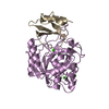
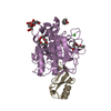
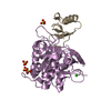
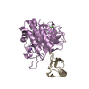
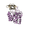
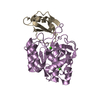
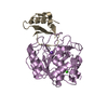
| Title | CALCIUM BINDING TO THERMITASE. CRYSTALLOGRAPHIC STUDIES OF THERMITASE AT 0, 5 AND 100 MM CALCIUM | ||||||
 Components Components |
| ||||||
 Keywords Keywords | COMPLEX(SERINE PROTEINASE-INHIBITOR) | ||||||
| Function / homology |  Function and homology information Function and homology informationthermitase / serine-type endopeptidase inhibitor activity / response to wounding / serine-type endopeptidase activity / proteolysis / extracellular region / metal ion binding Similarity search - Function | ||||||
| Biological species |  Thermoactinomyces vulgaris (bacteria) Thermoactinomyces vulgaris (bacteria) Hirudinaria manillensis (invertebrata) Hirudinaria manillensis (invertebrata) | ||||||
| Method |  X-RAY DIFFRACTION / Resolution: 2 Å X-RAY DIFFRACTION / Resolution: 2 Å | ||||||
 Authors Authors | Gros, P. / Kalk, K.H. / Hol, W.G.J. | ||||||
 Citation Citation | Journal: J.Biol.Chem. / Year: 1991 Title: Calcium binding to thermitase. Crystallographic studies of thermitase at 0, 5, and 100 mM calcium. Authors: Gros, P. / Kalk, K.H. / Hol, W.G. #1: Journal: Proteins / Year: 1992Title: Effects of Eglin-C Binding to Thermitase: Three-Dimensional Structure Comparison of Native Thermitase and Thermitase Eglin-C Complexes Authors: Gros, P. / Teplyakov, A.V. / Hol, W.G.J. | ||||||
| History |
|
- Structure visualization
| Structure viewer | Molecule: MolmilJmol/JSmol |
|---|
- Downloads & links
-Download
| PDBx/mmCIF format | 3tec.cif.gz | 74.3 KB | Display | PDBx/mmCIF format |
|---|---|---|---|---|
| PDB format | pdb3tec.ent.gz | 52.8 KB | Display | PDB format |
| PDBx/mmJSON format | 3tec.json.gz | Tree view | PDBx/mmJSON format | |
| Others |  Other downloads Other downloads |
-Validation report
| Arichive directory | https://data.pdbj.org/pub/pdb/validation_reports/te/3tecftp://data.pdbj.org/pub/pdb/validation_reports/te/3tec | HTTPS FTP |
|---|
-Related structure data
| Similar structure data |
|---|


-Links
 PDBj
PDBj

- Assembly
| Deposited unit | 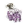
| ||||||||
|---|---|---|---|---|---|---|---|---|---|
| 1 |
| ||||||||
| Unit cell |
| ||||||||
| Atom site foot note | 1: RESIDUE PRO E 172 IS A CIS PROLINE. 2: THE BOND PRO E 214 - THR E 215 IS IN THE CIS CONFORMATION. |
-Components
| #1: Protein | Mass: 28384.896 Da / Num. of mol.: 1 Source method: isolated from a genetically manipulated source Source: (gene. exp.) Thermoactinomyces vulgaris (bacteria) / References: UniProt: P04072, thermitase | ||||
|---|---|---|---|---|---|
| #2: Protein | Mass: 8100.011 Da / Num. of mol.: 1 Source method: isolated from a genetically manipulated source Source: (gene. exp.) Hirudinaria manillensis (invertebrata) / Production host: unidentified (others) / References: UniProt: P01051 | ||||
| #3: Chemical |   Mass: 40.078 Da / Num. of mol.: 3 / Source method: obtained synthetically / Formula: Ca Mass: 40.078 Da / Num. of mol.: 3 / Source method: obtained synthetically / Formula: Ca#4: Water | ChemComp-HOH / |  Mass: 18.015 Da / Num. of mol.: 130 / Source method: isolated from a natural source / Formula: H2O Mass: 18.015 Da / Num. of mol.: 130 / Source method: isolated from a natural source / Formula: H2OSequence details | THERMITASE RESIDUE 199 IS A VALINE ACCORDING TO AMINO ACID SEQUENCE DETERMINATION BY MELOUN ET AL. ...THERMITASE | |
-Experimental details
-Experiment
| Experiment | Method: X-RAY DIFFRACTION |
|---|
- Sample preparation
| Crystal | Density Matthews: 2.85 Å3/Da / Density % sol: 56.9 % | ||||||||||||||||||||||||||||||||||||
|---|---|---|---|---|---|---|---|---|---|---|---|---|---|---|---|---|---|---|---|---|---|---|---|---|---|---|---|---|---|---|---|---|---|---|---|---|---|
| Crystal grow | *PLUS Method: vapor diffusion, hanging drop / pH: 6 | ||||||||||||||||||||||||||||||||||||
| Components of the solutions | *PLUS
|
-Data collection
| Reflection | *PLUS Highest resolution: 2 Å / Num. obs: 22550 / % possible obs: 78 % / Observed criterion σ(I): 1 / Num. measured all: 91303 / Rmerge(I) obs: 0.066 |
|---|
- Processing
| Software | Name: GROMOS / Classification: refinement | ||||||||||||||||||||||||||||||||||||||||||||||||||||||||||||
|---|---|---|---|---|---|---|---|---|---|---|---|---|---|---|---|---|---|---|---|---|---|---|---|---|---|---|---|---|---|---|---|---|---|---|---|---|---|---|---|---|---|---|---|---|---|---|---|---|---|---|---|---|---|---|---|---|---|---|---|---|---|
| Refinement | Rfactor Rwork: 0.168 / Highest resolution: 2 Å | ||||||||||||||||||||||||||||||||||||||||||||||||||||||||||||
| Refinement step | Cycle: LAST / Highest resolution: 2 Å
| ||||||||||||||||||||||||||||||||||||||||||||||||||||||||||||
| Refine LS restraints |
| ||||||||||||||||||||||||||||||||||||||||||||||||||||||||||||
| Refinement | *PLUS Highest resolution: 2 Å / Rfactor obs: 0.168 | ||||||||||||||||||||||||||||||||||||||||||||||||||||||||||||
| Solvent computation | *PLUS | ||||||||||||||||||||||||||||||||||||||||||||||||||||||||||||
| Displacement parameters | *PLUS | ||||||||||||||||||||||||||||||||||||||||||||||||||||||||||||
| Refine LS restraints | *PLUS
|
