 Movie
Movie Controller


+ Open data

- Basic information
| Entry | Database: PDB / ID: 6u9l | |||||||||
|---|---|---|---|---|---|---|---|---|---|---|
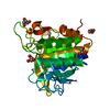
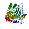
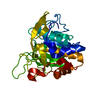
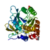
| Title | Imidazole-triggered RAS-specific subtilisin SUBT_BACAM | |||||||||
 Components Components | SUBTILISIN BPN' | |||||||||
 Keywords Keywords | HYDROLASE / engineered protease | |||||||||
| Function / homology | : / THIOCYANATE ION Function and homology information Function and homology information | |||||||||
| Biological species |  | |||||||||
| Method |  X-RAY DIFFRACTION / MOLECULAR REPLACEMENT / molecular replacement / Resolution: 1.7 Å X-RAY DIFFRACTION / MOLECULAR REPLACEMENT / molecular replacement / Resolution: 1.7 Å | |||||||||
 Authors Authors | Toth, E.A. / Bryan, P.N. / Orban, J. | |||||||||
| Funding support |  United States, 2items United States, 2items
| |||||||||
 Citation Citation | Journal: Commun Biol / Year: 2021 Title: Engineering subtilisin proteases that specifically degrade active RAS. Authors: Chen, Y. / Toth, E.A. / Ruan, B. / Choi, E.J. / Simmerman, R. / Chen, Y. / He, Y. / Wang, R. / Godoy-Ruiz, R. / King, H. / Custer, G. / Travis Gallagher, D. / Rozak, D.A. / Solomon, M. / ...Authors: Chen, Y. / Toth, E.A. / Ruan, B. / Choi, E.J. / Simmerman, R. / Chen, Y. / He, Y. / Wang, R. / Godoy-Ruiz, R. / King, H. / Custer, G. / Travis Gallagher, D. / Rozak, D.A. / Solomon, M. / Muro, S. / Weber, D.J. / Orban, J. / Fuerst, T.R. / Bryan, P.N. | |||||||||
| History |
|
- Structure visualization
| Structure viewer | Molecule: MolmilJmol/JSmol |
|---|
- Downloads & links
-Download
| PDBx/mmCIF format | 6u9l.cif.gz | 67.5 KB | Display | PDBx/mmCIF format |
|---|---|---|---|---|
| PDB format | pdb6u9l.ent.gz | 46.4 KB | Display | PDB format |
| PDBx/mmJSON format | 6u9l.json.gz | Tree view | PDBx/mmJSON format | |
| Others |  Other downloads Other downloads |
-Validation report
| Arichive directory | https://data.pdbj.org/pub/pdb/validation_reports/u9/6u9lftp://data.pdbj.org/pub/pdb/validation_reports/u9/6u9l | HTTPS FTP |
|---|
-Related structure data
| Related structure data |  6uaiC  6uaoC  6ubeC  1spbS S: Starting model for refinement C: citing same article ( |
|---|---|
| Similar structure data |


-Links
 PDBj
PDBj
- Assembly
| Deposited unit | 
| ||||||||
|---|---|---|---|---|---|---|---|---|---|
| 1 |
| ||||||||
| Unit cell |
|
-Components
| #1: Protein | Mass: 26456.518 Da / Num. of mol.: 1 Source method: isolated from a genetically manipulated source Source: (gene. exp.) | ||||||||||
|---|---|---|---|---|---|---|---|---|---|---|---|
| #2: Chemical |   Mass: 39.098 Da / Num. of mol.: 3 / Source method: obtained synthetically / Formula: K Mass: 39.098 Da / Num. of mol.: 3 / Source method: obtained synthetically / Formula: K#3: Chemical | ChemComp-GOL / |   Mass: 92.094 Da / Num. of mol.: 1 / Source method: obtained synthetically / Formula: C3H8O3 Mass: 92.094 Da / Num. of mol.: 1 / Source method: obtained synthetically / Formula: C3H8O3#4: Chemical | ChemComp-SCN /   Mass: 58.082 Da / Num. of mol.: 5 / Source method: obtained synthetically / Formula: CNS Mass: 58.082 Da / Num. of mol.: 5 / Source method: obtained synthetically / Formula: CNS#5: Water | ChemComp-HOH / |  Mass: 18.015 Da / Num. of mol.: 185 / Source method: isolated from a natural source / Formula: H2O Mass: 18.015 Da / Num. of mol.: 185 / Source method: isolated from a natural source / Formula: H2OHas ligand of interest | N | Has protein modification | Y | |
-Experimental details
-Experiment
| Experiment | Method: X-RAY DIFFRACTION / Number of used crystals: 1 |
|---|
- Sample preparation
| Crystal | Density Matthews: 2.03 Å3/Da / Density % sol: 39.33 % |
|---|---|
| Crystal grow | Temperature: 298 K / Method: vapor diffusion, sitting drop / pH: 8.5 Details: 0.1 M Bis-TRIS propane pH 8.5, 0.2 M KSCN, and 20% PEG 3350 |
-Data collection
| Diffraction | Mean temperature: 100 K / Serial crystal experiment: N |
|---|---|
| Diffraction source | Source: ROTATING ANODE / Type: RIGAKU MICROMAX-007 / Wavelength: 1.5418 Å |
| Detector | Type: RIGAKU RAXIS IV++ / Detector: IMAGE PLATE / Date: Aug 6, 2016 |
| Radiation | Protocol: SINGLE WAVELENGTH / Monochromatic (M) / Laue (L): M / Scattering type: x-ray |
| Radiation wavelength | Wavelength: 1.5418 Å / Relative weight: 1 |
| Reflection | Resolution: 1.7→53.08 Å / Num. obs: 24845 / % possible obs: 100 % / Redundancy: 27.2 % / CC1/2: 0.999 / Rmerge(I) obs: 0.097 / Rpim(I) all: 0.019 / Rrim(I) all: 0.099 / Net I/σ(I): 26.7 / Num. measured all: 675806 / Scaling rejects: 825 |
| Reflection shell | Resolution: 1.7→1.73 Å / Redundancy: 26.6 % / Rmerge(I) obs: 0.416 / Num. measured all: 34830 / Num. unique obs: 1307 / CC1/2: 0.98 / Rpim(I) all: 0.082 / Rrim(I) all: 0.424 / Net I/σ(I) obs: 8.3 / % possible all: 100 |
-Phasing
| Phasing | Method: molecular replacement | ||||||
|---|---|---|---|---|---|---|---|
| Phasing MR | R rigid body: 0.466
|
- Processing
| Software |
| ||||||||||||||||||||||||||||||||||||||||||||||||||||||||||||
|---|---|---|---|---|---|---|---|---|---|---|---|---|---|---|---|---|---|---|---|---|---|---|---|---|---|---|---|---|---|---|---|---|---|---|---|---|---|---|---|---|---|---|---|---|---|---|---|---|---|---|---|---|---|---|---|---|---|---|---|---|---|
| Refinement | Method to determine structure: MOLECULAR REPLACEMENT Starting model: 1spb Resolution: 1.7→53.08 Å / Cor.coef. Fo:Fc: 0.967 / Cor.coef. Fo:Fc free: 0.954 / WRfactor Rfree: 0.1697 / WRfactor Rwork: 0.1458 / FOM work R set: 0.9065 / SU B: 1.499 / SU ML: 0.051 / SU R Cruickshank DPI: 0.0933 / SU Rfree: 0.0888 / Cross valid method: THROUGHOUT / σ(F): 0 / ESU R: 0.093 / ESU R Free: 0.089 / Stereochemistry target values: MAXIMUM LIKELIHOOD Details: HYDROGENS HAVE BEEN ADDED IN THE RIDING POSITIONS U VALUES : REFINED INDIVIDUALLY
| ||||||||||||||||||||||||||||||||||||||||||||||||||||||||||||
| Solvent computation | Ion probe radii: 0.8 Å / Shrinkage radii: 0.8 Å / VDW probe radii: 1.2 Å / Solvent model: MASK | ||||||||||||||||||||||||||||||||||||||||||||||||||||||||||||
| Displacement parameters | Biso max: 48.54 Å2 / Biso mean: 13.729 Å2 / Biso min: 7.76 Å2
| ||||||||||||||||||||||||||||||||||||||||||||||||||||||||||||
| Refinement step | Cycle: final / Resolution: 1.7→53.08 Å
| ||||||||||||||||||||||||||||||||||||||||||||||||||||||||||||
| Refine LS restraints |
| ||||||||||||||||||||||||||||||||||||||||||||||||||||||||||||
| LS refinement shell | Resolution: 1.7→1.744 Å / Rfactor Rfree error: 0 / Total num. of bins used: 20
|
