 Movie
Movie Controller


+ Open data

- Basic information
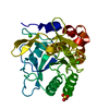
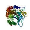
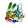
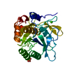
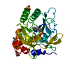
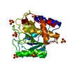
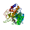
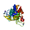
| Entry | Database: PDB / ID: 1c9m | ||||||
|---|---|---|---|---|---|---|---|
| Title | BACILLUS LENTUS SUBTILSIN (SER 87) N76D/S103A/V104I | ||||||
 Components Components | SERINE PROTEASE | ||||||
 Keywords Keywords | HYDROLASE / SERINE PROTEASE / SUBTILISIN / SITE-SPECIFIC VARIANT / ALTERED FLEXIBILITY | ||||||
| Function / homology |  Function and homology information Function and homology informationsubtilisin / sporulation resulting in formation of a cellular spore / serine-type endopeptidase activity / proteolysis / extracellular region / metal ion binding Similarity search - Function | ||||||
| Biological species |  Bacillus lentus (bacteria) Bacillus lentus (bacteria) | ||||||
| Method |  X-RAY DIFFRACTION / Resolution: 1.67 Å X-RAY DIFFRACTION / Resolution: 1.67 Å | ||||||
 Authors Authors | Bott, R. | ||||||
 Citation Citation | Journal: J.Mol.Biol. / Year: 1999 Title: Engineered Bacillus lentus subtilisins having altered flexibility. Authors: Graycar, T. / Knapp, M. / Ganshaw, G. / Dauberman, J. / Bott, R. | ||||||
| History |
|
- Structure visualization
| Structure viewer | Molecule: MolmilJmol/JSmol |
|---|
- Downloads & links
-Download
| PDBx/mmCIF format | 1c9m.cif.gz | 63.7 KB | Display | PDBx/mmCIF format |
|---|---|---|---|---|
| PDB format | pdb1c9m.ent.gz | 45.2 KB | Display | PDB format |
| PDBx/mmJSON format | 1c9m.json.gz | Tree view | PDBx/mmJSON format | |
| Others |  Other downloads Other downloads |
-Validation report
| Arichive directory | https://data.pdbj.org/pub/pdb/validation_reports/c9/1c9mftp://data.pdbj.org/pub/pdb/validation_reports/c9/1c9m | HTTPS FTP |
|---|
-Related structure data


-Links
 PDBj
PDBj

- Assembly
| Deposited unit | 
| ||||||||
|---|---|---|---|---|---|---|---|---|---|
| 1 |
| ||||||||
| Unit cell |
|
-Components
| #1: Protein | Mass: 26871.576 Da / Num. of mol.: 1 / Mutation: N76D, S103A, V104I FROM NATIVE (SER 87) Source method: isolated from a genetically manipulated source Source: (gene. exp.) Bacillus lentus (bacteria) / Production host: | ||||
|---|---|---|---|---|---|
| #2: Chemical | ChemComp-SO4 /   Mass: 96.063 Da / Num. of mol.: 1 / Source method: obtained synthetically / Formula: SO4 Mass: 96.063 Da / Num. of mol.: 1 / Source method: obtained synthetically / Formula: SO4 | ||||
| #3: Chemical |   Mass: 40.078 Da / Num. of mol.: 2 / Source method: obtained synthetically / Formula: Ca Mass: 40.078 Da / Num. of mol.: 2 / Source method: obtained synthetically / Formula: Ca#4: Water | ChemComp-HOH / |  Mass: 18.015 Da / Num. of mol.: 104 / Source method: isolated from a natural source / Formula: H2O Mass: 18.015 Da / Num. of mol.: 104 / Source method: isolated from a natural source / Formula: H2OHas protein modification | Y | |
-Experimental details
-Experiment
| Experiment | Method: X-RAY DIFFRACTION / Number of used crystals: 2 |
|---|
- Sample preparation
| Crystal | Density Matthews: 2.28 Å3/Da / Density % sol: 45.96 % | ||||||||||||||||||||||||||||||||||||
|---|---|---|---|---|---|---|---|---|---|---|---|---|---|---|---|---|---|---|---|---|---|---|---|---|---|---|---|---|---|---|---|---|---|---|---|---|---|
| Crystal grow | Method: vapor diffusion, hanging drop / pH: 5.9 Details: SODIUM ACTETATE, CALCIUM CHLORIDE, AMMONIUM SULFATE ,PHENYLMETHYLSULFANYL- FLOURIDE, pH 5.9, VAPOR DIFFUSION, HANGING DROP | ||||||||||||||||||||||||||||||||||||
| Crystal grow | *PLUS Method: vapor diffusion | ||||||||||||||||||||||||||||||||||||
| Components of the solutions | *PLUS
|
-Data collection
| Diffraction source | Source: ROTATING ANODE / Type: RIGAKU RU200 / Wavelength: 1.5418 |
|---|---|
| Detector | Type: RIGAKU RAXIS II / Detector: IMAGE PLATE / Date: Sep 10, 1993 |
| Radiation | Protocol: SINGLE WAVELENGTH / Monochromatic (M) / Laue (L): M / Scattering type: x-ray |
| Radiation wavelength | Wavelength: 1.5418 Å / Relative weight: 1 |
| Reflection | Resolution: 1.67→10 Å / Num. all: 26797 / Num. obs: 26797 / % possible obs: 91 % / Observed criterion σ(F): 2 / Redundancy: 2.5 % / Rmerge(I) obs: 0.04 |
| Reflection shell | Resolution: 1.67→10 Å / Redundancy: 2.5 % / Rmerge(I) obs: 0.04 / % possible all: 91 |
| Reflection | *PLUS Rmerge(I) obs: 0.04 |
- Processing
| Software | Name: PROLSQ / Classification: refinement | |||||||||||||||||||||||||||||||||||||||||||||||||||||||||||||||
|---|---|---|---|---|---|---|---|---|---|---|---|---|---|---|---|---|---|---|---|---|---|---|---|---|---|---|---|---|---|---|---|---|---|---|---|---|---|---|---|---|---|---|---|---|---|---|---|---|---|---|---|---|---|---|---|---|---|---|---|---|---|---|---|---|
| Refinement | Resolution: 1.67→10 Å / σ(F): 2
| |||||||||||||||||||||||||||||||||||||||||||||||||||||||||||||||
| Refinement step | Cycle: LAST / Resolution: 1.67→10 Å
| |||||||||||||||||||||||||||||||||||||||||||||||||||||||||||||||
| Refine LS restraints |
| |||||||||||||||||||||||||||||||||||||||||||||||||||||||||||||||
| Software | *PLUS Name: PROLSQ / Classification: refinement | |||||||||||||||||||||||||||||||||||||||||||||||||||||||||||||||
| Refinement | *PLUS σ(F): 2 | |||||||||||||||||||||||||||||||||||||||||||||||||||||||||||||||
| Solvent computation | *PLUS | |||||||||||||||||||||||||||||||||||||||||||||||||||||||||||||||
| Displacement parameters | *PLUS | |||||||||||||||||||||||||||||||||||||||||||||||||||||||||||||||
| Refine LS restraints | *PLUS
|
