 Movie
Movie Controller

[English] 日本語
 Yorodumi
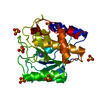
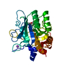
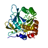
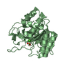
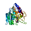
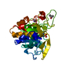
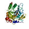
- PDB-1bh6: SUBTILISIN DY IN COMPLEX WITH THE SYNTHETIC INHIBITOR N-BENZYLOXY... -
+ Open data

- Basic information
| Entry | Database: PDB / ID: 1bh6 | ||||||
|---|---|---|---|---|---|---|---|
| Title | SUBTILISIN DY IN COMPLEX WITH THE SYNTHETIC INHIBITOR N-BENZYLOXYCARBONYL-ALA-PRO-PHE-CHLOROMETHYL KETONE | ||||||
 Components Components | SUBTILISIN DY | ||||||
 Keywords Keywords | HYDROLASE / SUBTILISIN / PROTEIN DEGRADATION | ||||||
| Function / homology |  Function and homology information Function and homology informationsubtilisin / sporulation resulting in formation of a cellular spore / serine-type endopeptidase activity / proteolysis / extracellular region / metal ion binding Similarity search - Function | ||||||
| Biological species |  | ||||||
| Method |  X-RAY DIFFRACTION / SYNCHROTRON / MOLECULAR REPLACEMENT / Resolution: 1.75 Å X-RAY DIFFRACTION / SYNCHROTRON / MOLECULAR REPLACEMENT / Resolution: 1.75 Å | ||||||
 Authors Authors | Eschenburg, S. / Genov, N. / Wilson, K.S. / Betzel, C. | ||||||
 Citation Citation | Journal: Eur.J.Biochem. / Year: 1998 Title: Crystal structure of subtilisin DY, a random mutant of subtilisin Carlsberg. Authors: Eschenburg, S. / Genov, N. / Peters, K. / Fittkau, S. / Stoeva, S. / Wilson, K.S. / Betzel, C. #1: Journal: Arch.Biochem.Biophys. / Year: 1993Title: Crystallization and Preliminary X-Ray Analysis of Subtilisin Dy, a Natural Mutant of Subtilisin Carlsberg Authors: Betzel, C. / Visanji, M. / Eschenburg, S. / Wilson, K.S. / Peters, K. / Fittkau, S. / Singh, T.P. / Genov, N. | ||||||
| History |
|
- Structure visualization
| Structure viewer | Molecule: MolmilJmol/JSmol |
|---|
- Downloads & links
-Download
| PDBx/mmCIF format | 1bh6.cif.gz | 69.5 KB | Display | PDBx/mmCIF format |
|---|---|---|---|---|
| PDB format | pdb1bh6.ent.gz | 49.6 KB | Display | PDB format |
| PDBx/mmJSON format | 1bh6.json.gz | Tree view | PDBx/mmJSON format | |
| Others |  Other downloads Other downloads |
-Validation report
| Arichive directory | https://data.pdbj.org/pub/pdb/validation_reports/bh/1bh6ftp://data.pdbj.org/pub/pdb/validation_reports/bh/1bh6 | HTTPS FTP |
|---|
-Related structure data
| Related structure data | 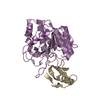 1cseS S: Starting model for refinement |
|---|---|
| Similar structure data |


-Links
 PDBj
PDBj

- Assembly
| Deposited unit | 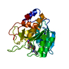
| ||||||||
|---|---|---|---|---|---|---|---|---|---|
| 1 |
| ||||||||
| Unit cell |
|
-Components
| #1: Protein | Mass: 27499.416 Da / Num. of mol.: 1 / Source method: isolated from a natural source Details: X-IRRADIATED JAPANESE STRAIN OF BACILLUS LICHENIFORMIS Source: (natural) |
|---|---|
| #2: Chemical | ChemComp-CA /   Mass: 40.078 Da / Num. of mol.: 1 / Source method: obtained synthetically / Formula: Ca Mass: 40.078 Da / Num. of mol.: 1 / Source method: obtained synthetically / Formula: Ca |
| #3: Chemical | ChemComp-NA /   Mass: 22.990 Da / Num. of mol.: 1 / Source method: obtained synthetically / Formula: Na Mass: 22.990 Da / Num. of mol.: 1 / Source method: obtained synthetically / Formula: Na |
| #4: Chemical | ChemComp-1BH /   Mass: 467.557 Da / Num. of mol.: 1 / Source method: obtained synthetically / Formula: C26H33N3O5 Mass: 467.557 Da / Num. of mol.: 1 / Source method: obtained synthetically / Formula: C26H33N3O5 |
| #5: Water | ChemComp-HOH /  Mass: 18.015 Da / Num. of mol.: 226 / Source method: isolated from a natural source / Formula: H2O Mass: 18.015 Da / Num. of mol.: 226 / Source method: isolated from a natural source / Formula: H2O |
| Has protein modification | Y |
-Experimental details
-Experiment
| Experiment | Method: X-RAY DIFFRACTION / Number of used crystals: 1 |
|---|
- Sample preparation
| Crystal | Density Matthews: 2.1 Å3/Da / Density % sol: 41 % | ||||||||||||||||||||||||||||||||||||||||||||||||||||||||||||
|---|---|---|---|---|---|---|---|---|---|---|---|---|---|---|---|---|---|---|---|---|---|---|---|---|---|---|---|---|---|---|---|---|---|---|---|---|---|---|---|---|---|---|---|---|---|---|---|---|---|---|---|---|---|---|---|---|---|---|---|---|---|
| Crystal grow | pH: 6 / Details: AS GIVEN IN REFERENCE 1, pH 6.0 | ||||||||||||||||||||||||||||||||||||||||||||||||||||||||||||
| Crystal | *PLUS | ||||||||||||||||||||||||||||||||||||||||||||||||||||||||||||
| Crystal grow | *PLUS Temperature: 16 ℃ / Method: vapor diffusion, hanging drop | ||||||||||||||||||||||||||||||||||||||||||||||||||||||||||||
| Components of the solutions | *PLUS
|
-Data collection
| Diffraction | Mean temperature: 278 K |
|---|---|
| Diffraction source | Source: SYNCHROTRON / Site: EMBL/DESY, HAMBURG  / Beamline: X11 / Wavelength: 0.92 / Beamline: X11 / Wavelength: 0.92 |
| Detector | Type: MARRESEARCH / Detector: IMAGE PLATE / Date: Feb 1, 1993 |
| Radiation | Monochromatic (M) / Laue (L): M / Scattering type: x-ray |
| Radiation wavelength | Wavelength: 0.92 Å / Relative weight: 1 |
| Reflection | Resolution: 1.75→10 Å / Num. obs: 22887 / % possible obs: 96.7 % / Observed criterion σ(I): 0 / Redundancy: 4.4 % / Rmerge(I) obs: 0.03 / Rsym value: 0.03 / Net I/σ(I): 23 |
| Reflection shell | Resolution: 1.75→1.79 Å / Redundancy: 3.1 % / Rmerge(I) obs: 0.052 / Mean I/σ(I) obs: 14 / Rsym value: 0.052 / % possible all: 92.2 |
| Reflection | *PLUS Num. measured all: 101587 |
| Reflection shell | *PLUS % possible obs: 92.2 % |
- Processing
| Software |
| ||||||||||||||||||||||||||||||||||||||||||||||||||||||||||||||||||||||||||||||||||||
|---|---|---|---|---|---|---|---|---|---|---|---|---|---|---|---|---|---|---|---|---|---|---|---|---|---|---|---|---|---|---|---|---|---|---|---|---|---|---|---|---|---|---|---|---|---|---|---|---|---|---|---|---|---|---|---|---|---|---|---|---|---|---|---|---|---|---|---|---|---|---|---|---|---|---|---|---|---|---|---|---|---|---|---|---|---|
| Refinement | Method to determine structure: MOLECULAR REPLACEMENT Starting model: PDB ENTRY 1CSE Resolution: 1.75→10 Å / σ(F): 0 /
| ||||||||||||||||||||||||||||||||||||||||||||||||||||||||||||||||||||||||||||||||||||
| Refine analyze | Luzzati sigma a obs: 0.12 Å | ||||||||||||||||||||||||||||||||||||||||||||||||||||||||||||||||||||||||||||||||||||
| Refinement step | Cycle: LAST / Resolution: 1.75→10 Å
| ||||||||||||||||||||||||||||||||||||||||||||||||||||||||||||||||||||||||||||||||||||
| Refine LS restraints |
|
