 Movie
Movie Controller


+ Open data

- Basic information
| Entry | Database: PDB / ID: 7am3 | ||||||
|---|---|---|---|---|---|---|---|
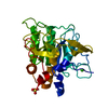
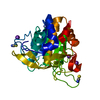
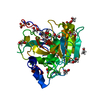
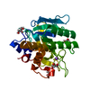
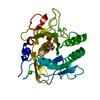
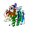
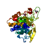
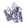
| Title | Crystal structure of Peptiligase mutant - M222P | ||||||
 Components Components | Subtilisin BPN' | ||||||
 Keywords Keywords | LIGASE / subtilisin / peptide ligase | ||||||
| Function / homology |  Function and homology information Function and homology informationsubtilisin / sporulation resulting in formation of a cellular spore / fibrinolysis / serine-type endopeptidase activity / proteolysis / extracellular region / metal ion binding Similarity search - Function | ||||||
| Biological species |  | ||||||
| Method |  X-RAY DIFFRACTION / MOLECULAR REPLACEMENT / Resolution: 1.61 Å X-RAY DIFFRACTION / MOLECULAR REPLACEMENT / Resolution: 1.61 Å | ||||||
 Authors Authors | Rozeboom, H.J. / Janssen, D.J. | ||||||
 Citation Citation | Journal: Comput Struct Biotechnol J / Year: 2021 Title: From thiol-subtilisin to omniligase: Design and structure of a broadly applicable peptide ligase. Authors: Toplak, A. / Teixeira de Oliveira, E.F. / Schmidt, M. / Rozeboom, H.J. / Wijma, H.J. / Meekels, L.K.M. / de Visser, R. / Janssen, D.B. / Nuijens, T. | ||||||
| History |
|
- Structure visualization
| Structure viewer | Molecule: MolmilJmol/JSmol |
|---|
- Downloads & links
-Download
| PDBx/mmCIF format | 7am3.cif.gz | 115.2 KB | Display | PDBx/mmCIF format |
|---|---|---|---|---|
| PDB format | pdb7am3.ent.gz | 86.9 KB | Display | PDB format |
| PDBx/mmJSON format | 7am3.json.gz | Tree view | PDBx/mmJSON format | |
| Others |  Other downloads Other downloads |
-Validation report
| Arichive directory | https://data.pdbj.org/pub/pdb/validation_reports/am/7am3ftp://data.pdbj.org/pub/pdb/validation_reports/am/7am3 | HTTPS FTP |
|---|
-Related structure data
| Related structure data | 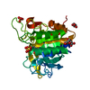 7am4C 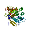 7am5C 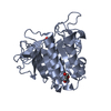 7am6C 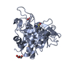 7am7C 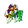 7am8C 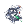 5ox2S S: Starting model for refinement C: citing same article ( |
|---|---|
| Similar structure data |


-Links
 PDBj
PDBj

- Assembly
| Deposited unit | 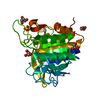
| ||||||||
|---|---|---|---|---|---|---|---|---|---|
| 1 |
| ||||||||
| Unit cell |
| ||||||||
| Components on special symmetry positions |
|
-Components
| #1: Protein | Mass: 27416.408 Da / Num. of mol.: 1 Mutation: Q2K S3C P5S S9A I31L S212C P216A M50F A73L DELTA75-83 E156S G166S G169A S188P Q206C N212G K217L N218S S221A T254A Q271E M222P Source method: isolated from a genetically manipulated source Source: (gene. exp.) | ||||||||
|---|---|---|---|---|---|---|---|---|---|
| #2: Chemical | ChemComp-GOL /   Mass: 92.094 Da / Num. of mol.: 4 / Source method: obtained synthetically / Formula: C3H8O3 Mass: 92.094 Da / Num. of mol.: 4 / Source method: obtained synthetically / Formula: C3H8O3#3: Chemical |   Mass: 96.063 Da / Num. of mol.: 2 / Source method: obtained synthetically / Formula: SO4 Mass: 96.063 Da / Num. of mol.: 2 / Source method: obtained synthetically / Formula: SO4#4: Water | ChemComp-HOH / |  Mass: 18.015 Da / Num. of mol.: 323 / Source method: isolated from a natural source / Formula: H2O Mass: 18.015 Da / Num. of mol.: 323 / Source method: isolated from a natural source / Formula: H2OHas ligand of interest | N | Has protein modification | Y | |
-Experimental details
-Experiment
| Experiment | Method: X-RAY DIFFRACTION / Number of used crystals: 1 |
|---|
- Sample preparation
| Crystal | Density Matthews: 1.8 Å3/Da / Density % sol: 37.42 % |
|---|---|
| Crystal grow | Temperature: 294 K / Method: vapor diffusion, hanging drop / pH: 6.5 / Details: 1.4 M MgSO4 , 0.1 M MES pH 6.5. |
-Data collection
| Diffraction | Mean temperature: 100 K / Serial crystal experiment: N |
|---|---|
| Diffraction source | Source: ROTATING ANODE / Type: BRUKER AXS MICROSTAR-H / Wavelength: 1.5418 Å |
| Detector | Type: MARRESEARCH / Detector: IMAGE PLATE / Date: Jan 7, 2019 |
| Radiation | Monochromator: HELIOS MX MIRRORS / Protocol: SINGLE WAVELENGTH / Monochromatic (M) / Laue (L): M / Scattering type: x-ray |
| Radiation wavelength | Wavelength: 1.5418 Å / Relative weight: 1 |
| Reflection | Resolution: 1.61→42.86 Å / Num. obs: 29222 / % possible obs: 99.6 % / Redundancy: 7.6 % / CC1/2: 0.999 / Rmerge(I) obs: 0.05 / Rpim(I) all: 0.019 / Rrim(I) all: 0.054 / Net I/σ(I): 26 |
| Reflection shell | Resolution: 1.61→1.64 Å / Redundancy: 6.6 % / Rmerge(I) obs: 0.197 / Num. unique obs: 1315 / CC1/2: 0.982 / Rpim(I) all: 0.081 / Rrim(I) all: 0.214 / % possible all: 91.6 |
- Processing
| Software |
| ||||||||||||||||||||||||||||||||||||||||||||||||||||||||||||
|---|---|---|---|---|---|---|---|---|---|---|---|---|---|---|---|---|---|---|---|---|---|---|---|---|---|---|---|---|---|---|---|---|---|---|---|---|---|---|---|---|---|---|---|---|---|---|---|---|---|---|---|---|---|---|---|---|---|---|---|---|---|
| Refinement | Method to determine structure: MOLECULAR REPLACEMENT Starting model: 5OX2 Resolution: 1.61→42.86 Å / Cor.coef. Fo:Fc: 0.972 / Cor.coef. Fo:Fc free: 0.962 / SU B: 2.286 / SU ML: 0.042 / Cross valid method: THROUGHOUT / σ(F): 0 / ESU R: 0.078 / ESU R Free: 0.075 / Stereochemistry target values: MAXIMUM LIKELIHOOD Details: U VALUES : WITH TLS ADDED HYDROGENS HAVE BEEN ADDED IN THE RIDING POSITIONS
| ||||||||||||||||||||||||||||||||||||||||||||||||||||||||||||
| Solvent computation | Ion probe radii: 0.8 Å / Shrinkage radii: 0.8 Å / VDW probe radii: 1.2 Å / Solvent model: MASK | ||||||||||||||||||||||||||||||||||||||||||||||||||||||||||||
| Displacement parameters | Biso max: 59.08 Å2 / Biso mean: 13.749 Å2 / Biso min: 5.85 Å2
| ||||||||||||||||||||||||||||||||||||||||||||||||||||||||||||
| Refinement step | Cycle: final / Resolution: 1.61→42.86 Å
| ||||||||||||||||||||||||||||||||||||||||||||||||||||||||||||
| Refine LS restraints |
| ||||||||||||||||||||||||||||||||||||||||||||||||||||||||||||
| LS refinement shell | Resolution: 1.61→1.65 Å / Rfactor Rfree error: 0
| ||||||||||||||||||||||||||||||||||||||||||||||||||||||||||||
| Refinement TLS params. | Method: refined / Origin x: 14.93 Å / Origin y: -12.704 Å / Origin z: 4.107 Å
|
