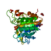
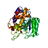
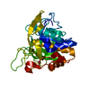
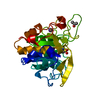
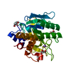
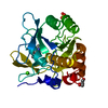
- PDB-7am6: Crystal structure of Peptiligase mutant - L217H/M222P/A225N/F189W -
+
Open data
ID or keywords:
Loading...
-
Basic information
Entry
Database: PDB / ID: 7am6
Title
Crystal structure of Peptiligase mutant - L217H/M222P/A225N/F189W
Components
(Subtilisin BPN') x 2
LEU-PRO-GLU-GLY-SER-PRO-VAL-THR-ASP-LEU-ARG-TYR
Keywords
LIGASE / subtilisin / peptide ligase
Function / homology
Function and homology information
subtilisin / sporulation resulting in formation of a cellular spore / fibrinolysis / serine-type endopeptidase inhibitor activity / response to wounding / serine-type endopeptidase activity / proteolysis / extracellular region / metal ion binding Similarity search - Function
Proteinase inhibitor I13, potato inhibitor I / Proteinase inhibitor I13, potato inhibitor I superfamily / Potato inhibitor I family / Potato inhibitor I family signature. / : / Fervidolysin N-terminal prodomain / Subtilisin Carlsberg-like catalytic domain / Peptidase S8 propeptide/proteinase inhibitor I9 superfamily / Peptidase S8/S53 domain / Peptidase S8, subtilisin, His-active site ...Proteinase inhibitor I13, potato inhibitor I / Proteinase inhibitor I13, potato inhibitor I superfamily / Potato inhibitor I family / Potato inhibitor I family signature. / : / Fervidolysin N-terminal prodomain / Subtilisin Carlsberg-like catalytic domain / Peptidase S8 propeptide/proteinase inhibitor I9 superfamily / Peptidase S8/S53 domain / Peptidase S8, subtilisin, His-active site / : / Serine proteases, subtilase family, histidine active site. / Serine proteases, subtilase family, aspartic acid active site. / Peptidase S8, subtilisin, Asp-active site / Serine proteases, subtilase family, serine active site. / Peptidase S8, subtilisin, Ser-active site / Peptidase S8, subtilisin-related / Serine proteases, subtilase domain profile. / Peptidase S8/S53 domain superfamily / Subtilase family / Peptidase S8/S53 domain / Rossmann fold / 3-Layer(aba) Sandwich / Alpha Beta Similarity search - Domain/homology
In the structure databanks used in Yorodumi, some data are registered as the other names, "COVID-19 virus" and "2019-nCoV". Here are the details of the virus and the list of structure data.
Jan 31, 2019. EMDB accession codes are about to change! (news from PDBe EMDB page)
EMDB accession codes are about to change! (news from PDBe EMDB page)
The allocation of 4 digits for EMDB accession codes will soon come to an end. Whilst these codes will remain in use, new EMDB accession codes will include an additional digit and will expand incrementally as the available range of codes is exhausted. The current 4-digit format prefixed with “EMD-” (i.e. EMD-XXXX) will advance to a 5-digit format (i.e. EMD-XXXXX), and so on. It is currently estimated that the 4-digit codes will be depleted around Spring 2019, at which point the 5-digit format will come into force.
The EM Navigator/Yorodumi systems omit the EMD- prefix.
Related info.:Q: What is EMD? / ID/Accession-code notation in Yorodumi/EM Navigator
Yorodumi is a browser for structure data from EMDB, PDB, SASBDB, etc.
This page is also the successor to EM Navigator detail page, and also detail information page/front-end page for Omokage search.
The word "yorodu" (or yorozu) is an old Japanese word meaning "ten thousand". "mi" (miru) is to see.
Related info.:EMDB / PDB / SASBDB / Comparison of 3 databanks / Yorodumi Search / Aug 31, 2016. New EM Navigator & Yorodumi / Yorodumi Papers / Jmol/JSmol / Function and homology information / Changes in new EM Navigator and Yorodumi
 Movie
Movie Controller

 Yorodumi
 Open data
 Basic information
 Components
 Keywords
 Function and homology information
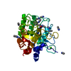
 Hirudo medicinalis (medicinal leech)
 X-RAY DIFFRACTION /
  Authors
 Citation
 Structure visualization
 Downloads & links
 Other downloads
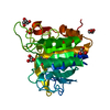


 PDBj
PDBj


 Assembly


 Mass: 92.094 Da / Num. of mol.: 8 / Source method: obtained synthetically / Formula: C3H8O3
 Mass: 150.087 Da / Num. of mol.: 2 / Source method: obtained synthetically / Formula: C4H6O6
 Sample preparation
 Processing