 Movie
Movie Controller


+ Open data

- Basic information
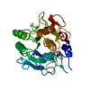
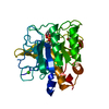
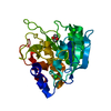
| Entry | Database: PDB / ID: 1sbt | |||||||||
|---|---|---|---|---|---|---|---|---|---|---|
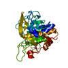
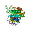
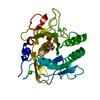
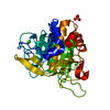
| Title | ATOMIC COORDINATES FOR SUBTILISIN BPN (OR NOVO) | |||||||||
 Components Components | SUBTILISIN BPN' | |||||||||
 Keywords Keywords | HYDROLASE (SERINE PROTEINASE) | |||||||||
| Function / homology |  Function and homology information Function and homology informationsubtilisin / sporulation resulting in formation of a cellular spore / fibrinolysis / serine-type endopeptidase activity / proteolysis / extracellular region / metal ion binding Similarity search - Function | |||||||||
| Biological species |  | |||||||||
| Method |  X-RAY DIFFRACTION / Resolution: 2.5 Å X-RAY DIFFRACTION / Resolution: 2.5 Å | |||||||||
 Authors Authors | Alden, R.A. / Birktoft, J.J. / Kraut, J. / Robertus, J.D. / Wright, C.S. | |||||||||
 Citation Citation | Journal: Biochem.Biophys.Res.Commun. / Year: 1971 Title: Atomic coordinates for subtilisin BPN' (or Novo). Authors: Alden, R.A. / Birktoft, J.J. / Kraut, J. / Robertus, J.D. / Wright, C.S. #1: Journal: J.Biol.Chem. / Year: 1976Title: Polypeptide Halomethyl Ketones Bind to Serine Proteases as Analogs of the Tetrahedral Intermediate,X-Ray Crystallographic Comparison of Lysine-and Phenylalanine-Polypeptide Chloromethyl Ketone-Inhibited Subtilisin Authors: Poulos, T.L. / Alden, R.A. / Freer, S.T. / Birktoft, J.J. / Kraut, J. #2: Journal: J.Biol.Chem. / Year: 1975Title: X-Ray Crystallographic Study of Boronic Acid Adducts with Subtilisin Bpn(Novo),A Model for the Catalytic Transition State Authors: Matthews, D.A. / Alden, R.A. / Birktoft, J.J. / Freer, S.T. / Kraut, J. #3: Journal: Biochemistry / Year: 1972Title: Subtilisin,A Stereochemical Mechanism Involving Transition-State Stabilization Authors: Robertus, J.D. / Kraut, J. / Alden, R.A. / Birktoft, J.J. #4: Journal: Biochemistry / Year: 1972Title: An X-Ray Crystallographic Study of the Binding of Peptide Chloromethyl Ketone Inhibitors to Subtilisin Bpn Authors: Robertus, J.D. / Alden, R.A. / Birktoft, J.J. / Kraut, J. / Powers, J.C. / Wilcox, P.E. #5: Journal: Cold Spring Harbor Symp.Quant.Biol. / Year: 1972Title: The Aromatic Substrate Binding Site in Subtilisin Bpnand its Resemblance to Chymotrypsin Authors: Kraut, J. / Robertus, J.D. / Birktoft, J.J. / Alden, R.A. / Wilcox, P.E. / Powers, J.C. #6: Journal: Biochem.Biophys.Res.Commun. / Year: 1971Title: On the Identity of Subtilisins Bpnand Novo Authors: Robertus, J.D. / Alden, R.A. / Kraut, J. #7: Journal: Philos.Trans.R.Soc.London,Ser.B / Year: 1970Title: A Hydrogen-Bond Network at the Active Site of Subtilisin Bpn Authors: Alden, R.A. / Wright, C.S. / Kraut, J. #8: Journal: Nature / Year: 1969Title: Structure of Subtilisin Bpnat 2.5 Angstroms Resolution Authors: Wright, C.S. / Alden, R.A. / Kraut, J. | |||||||||
| History |
|
- Structure visualization
| Structure viewer | Molecule: MolmilJmol/JSmol |
|---|
- Downloads & links
-Download
| PDBx/mmCIF format | 1sbt.cif.gz | 55.1 KB | Display | PDBx/mmCIF format |
|---|---|---|---|---|
| PDB format | pdb1sbt.ent.gz | 32.4 KB | Display | PDB format |
| PDBx/mmJSON format | 1sbt.json.gz | Tree view | PDBx/mmJSON format | |
| Others |  Other downloads Other downloads |
-Validation report
| Arichive directory | https://data.pdbj.org/pub/pdb/validation_reports/sb/1sbtftp://data.pdbj.org/pub/pdb/validation_reports/sb/1sbt | HTTPS FTP |
|---|
-Related structure data
| Similar structure data |
|---|


-Links
 PDBj
PDBj

- Assembly
| Deposited unit | 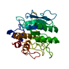
| ||||||||
|---|---|---|---|---|---|---|---|---|---|
| 1 |
| ||||||||
| Unit cell |
| ||||||||
| Atom site foot note | 1: THE FO-FC MAP INDICATES COORDINATE ERRORS OF MORE THAN 1 ANGSTROM. 2: THESE COORDINATES ARE THE OUTPUT OF A REFINEMENT BY DIAMOND'S MODEL-BUILDING PROGRAM. |
-Components
| #1: Protein | Mass: 27552.525 Da / Num. of mol.: 1 Source method: isolated from a genetically manipulated source Source: (gene. exp.) |
|---|---|
| #2: Water | ChemComp-HOH /  Mass: 18.015 Da / Num. of mol.: 17 / Source method: isolated from a natural source / Formula: H2O Mass: 18.015 Da / Num. of mol.: 17 / Source method: isolated from a natural source / Formula: H2O |
-Experimental details
-Experiment
| Experiment | Method: X-RAY DIFFRACTION |
|---|
- Sample preparation
| Crystal | Density Matthews: 2.07 Å3/Da / Density % sol: 40.54 % |
|---|---|
| Crystal grow | *PLUS Method: other / Details: Wright, C.S., (1969) Nature, 221, 235. |
- Processing
| Refinement | Highest resolution: 2.5 Å Details: THE FO-FC MAP INDICATES COORDINATE ERRORS OF MORE THAN 1 ANGSTROM. THESE COORDINATES ARE THE OUTPUT OF A REFINEMENT BY DIAMOND'S MODEL-BUILDING PROGRAM | ||||||||||||
|---|---|---|---|---|---|---|---|---|---|---|---|---|---|
| Refinement step | Cycle: LAST / Highest resolution: 2.5 Å
|
