- PDB-1sup: SUBTILISIN BPN' AT 1.6 ANGSTROMS RESOLUTION: ANALYSIS OF DISCRETE... -
+
Open data
ID or keywords:
Loading...
-
Basic information
Entry
Database: PDB / ID: 1sup
Title
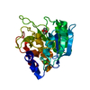
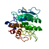
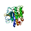
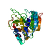
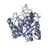
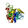
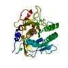
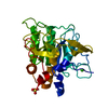
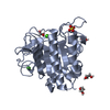
SUBTILISIN BPN' AT 1.6 ANGSTROMS RESOLUTION: ANALYSIS OF DISCRETE DISORDER AND COMPARISON OF CRYSTAL FORMS
Components
SUBTILISIN BPN'
Keywords
HYDROLASE (SERINE PROTEASE)
Function / homology
Function and homology information
subtilisin / sporulation resulting in formation of a cellular spore / fibrinolysis / serine-type endopeptidase activity / proteolysis / extracellular region / metal ion binding Similarity search - Function
Mass: 18.015 Da / Num. of mol.: 194 / Source method: isolated from a natural source / Formula: H2O
Compound details
SER 221 IS COVALENTLY DERIVATIZED WITH PHENYLMETHYLSULFONATE (PMS), HET GROUP 278.
Has protein modification
Y
-
Experimental details
-
Experiment
Experiment
Method: X-RAY DIFFRACTION
-
Sample preparation
Crystal
Density Matthews: 2.05 Å3/Da / Density % sol: 39.98 %
Crystal grow
*PLUS
pH: 7 / Method: vapor diffusion, hanging drop
Components of the solutions
*PLUS
ID
Conc.
Common name
Crystal-ID
Sol-ID
Details (eV)
1
7.5mg/ml
protein
1
drop
2
20mM
MES
1
drop
pH7.0
3
2mM
PMSF
1
drop
4
35-40 %saturated
ammoniumsulfate
1
reservoir
5
0.5 %
PEG8000
1
reservoir
6
1 %
beta-octylglucoside
1
reservoir
7
2 %
MPD
1
reservoir
8
50mM
MES
1
reservoir
pH7.0
-
Data collection
Reflection
Num. obs: 26328 / % possible obs: 92 %
-
Processing
Software
Name: PROLSQ / Classification: refinement
Refinement
Highest resolution: 1.6 Å / σ(F): 2 Details: PEPTIDE 257 - 258 IS DISCRETELY DISORDERED. THE ZONE 256 - 259 WAS REFINED IN TWO DISTINCT CONFORMATIONS. ATOMS CD, CE, AND NZ OF LYS 256 ARE FURTHER DISORDERED, SO THEIR OCCUPANCIES DO NOT SUM TO 1.0.
In the structure databanks used in Yorodumi, some data are registered as the other names, "COVID-19 virus" and "2019-nCoV". Here are the details of the virus and the list of structure data.
Jan 31, 2019. EMDB accession codes are about to change! (news from PDBe EMDB page)
EMDB accession codes are about to change! (news from PDBe EMDB page)
The allocation of 4 digits for EMDB accession codes will soon come to an end. Whilst these codes will remain in use, new EMDB accession codes will include an additional digit and will expand incrementally as the available range of codes is exhausted. The current 4-digit format prefixed with “EMD-” (i.e. EMD-XXXX) will advance to a 5-digit format (i.e. EMD-XXXXX), and so on. It is currently estimated that the 4-digit codes will be depleted around Spring 2019, at which point the 5-digit format will come into force.
The EM Navigator/Yorodumi systems omit the EMD- prefix.
Related info.:Q: What is EMD? / ID/Accession-code notation in Yorodumi/EM Navigator
Yorodumi is a browser for structure data from EMDB, PDB, SASBDB, etc.
This page is also the successor to EM Navigator detail page, and also detail information page/front-end page for Omokage search.
The word "yorodu" (or yorozu) is an old Japanese word meaning "ten thousand". "mi" (miru) is to see.
Related info.:EMDB / PDB / SASBDB / Comparison of 3 databanks / Yorodumi Search / Aug 31, 2016. New EM Navigator & Yorodumi / Yorodumi Papers / Jmol/JSmol / Function and homology information / Changes in new EM Navigator and Yorodumi
 Movie
Movie Controller

 Yorodumi
 Open data
 Basic information
 Components
 Keywords
 Function and homology information
 X-RAY DIFFRACTION / Resolution: 1.6 Å
  Authors
 Citation
 Structure visualization
 Downloads & links
 Other downloads
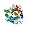
 PDBj
PDBj

 Assembly
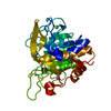

 Mass: 40.078 Da / Num. of mol.: 1 / Source method: obtained synthetically / Formula: Ca
 Mass: 22.990 Da / Num. of mol.: 1 / Source method: obtained synthetically / Formula: Na
 Mass: 172.202 Da / Num. of mol.: 1 / Source method: obtained synthetically / Formula: C7H8O3S
 Mass: 18.015 Da / Num. of mol.: 194 / Source method: isolated from a natural source / Formula: H2O
 Sample preparation
 Processing