[English] 日本語
Yorodumi
- PDB-4b72: Aminoimidazoles as BACE-1 Inhibitors: From De Novo Design to Ab- ... -

+
Open data


ID or keywords:

Loading...

-
Basic information

Entry
Database: PDB / ID: 4b72
TitleAminoimidazoles as BACE-1 Inhibitors: From De Novo Design to Ab- lowering in Brain
ComponentsBETA-SECRETASE 1
KeywordsHYDROLASE / HYDROLASE INHIBITOR / LEAD GENERATION / STRUCTURE-BASED DRUG DESIGN
Function / homology
Function and homology information


memapsin 2 / Golgi-associated vesicle lumen / signaling receptor ligand precursor processing / beta-aspartyl-peptidase activity / amyloid precursor protein catabolic process / amyloid-beta formation / membrane protein ectodomain proteolysis / cellular response to manganese ion / amyloid-beta metabolic process / prepulse inhibition ...memapsin 2 / Golgi-associated vesicle lumen / signaling receptor ligand precursor processing / beta-aspartyl-peptidase activity / amyloid precursor protein catabolic process / amyloid-beta formation / membrane protein ectodomain proteolysis / cellular response to manganese ion / amyloid-beta metabolic process / prepulse inhibition / detection of mechanical stimulus involved in sensory perception of pain / protein serine/threonine kinase binding / cellular response to copper ion / presynaptic modulation of chemical synaptic transmission / hippocampal mossy fiber to CA3 synapse / multivesicular body / response to lead ion / trans-Golgi network / protein processing / recycling endosome / cellular response to amyloid-beta / positive regulation of neuron apoptotic process / synaptic vesicle / late endosome / peptidase activity / amyloid-beta binding / endopeptidase activity / amyloid fibril formation / aspartic-type endopeptidase activity / lysosome / early endosome / endosome membrane / endosome / membrane raft / Amyloid fiber formation / endoplasmic reticulum lumen / axon / neuronal cell body / dendrite / Golgi apparatus / enzyme binding / cell surface / proteolysis / membrane / plasma membrane
Similarity search - Function
Beta-secretase BACE1 / Beta-secretase BACE / Memapsin-like / Eukaryotic aspartyl protease / Aspartic peptidase A1 family / Peptidase family A1 domain / Peptidase family A1 domain profile. / Cathepsin D, subunit A; domain 1 / Acid Proteases / Aspartic peptidase, active site ...Beta-secretase BACE1 / Beta-secretase BACE / Memapsin-like / Eukaryotic aspartyl protease / Aspartic peptidase A1 family / Peptidase family A1 domain / Peptidase family A1 domain profile. / Cathepsin D, subunit A; domain 1 / Acid Proteases / Aspartic peptidase, active site / Eukaryotic and viral aspartyl proteases active site. / Aspartic peptidase domain superfamily / Beta Barrel / Mainly Beta
Similarity search - Domain/homology
Chem-2FB / Beta-secretase 1
Similarity search - Component
Biological speciesHOMO SAPIENS (human)
MethodX-RAY DIFFRACTION / MOLECULAR REPLACEMENT / Resolution: 1.6 Å
AuthorsGravenfors, Y. / Blid, J. / Ginman, T. / Karlstrom, S. / Kihlstrom, J. / Kolmodin, K. / Lindstrom, J. / Berg, S. / Kieseritzky, F. / Slivo, C. ...Gravenfors, Y. / Blid, J. / Ginman, T. / Karlstrom, S. / Kihlstrom, J. / Kolmodin, K. / Lindstrom, J. / Berg, S. / Kieseritzky, F. / Slivo, C. / Swahn, B. / Viklund, J. / Olsson, L. / Johansson, P. / Eketjall, S. / Falting, J. / Jeppsson, F. / Stromberg, K. / Janson, J. / Rahm, F.
CitationJournal: J.Med.Chem. / Year: 2013
Title: Core Refinement Toward Permeable Beta-Secretase (Bace-1) Inhibitors with Low Herg Activity.
Authors: Ginman, T. / Viklund, J. / Malmstrom, J. / Blid, J. / Emond, R. / Forsblom, R. / Johansson, A. / Kers, A. / Lake, F. / Sehgelmeble, F. / Sterky, K.J. / Bergh, M. / Lindgren, A. / Johansson, ...Authors: Ginman, T. / Viklund, J. / Malmstrom, J. / Blid, J. / Emond, R. / Forsblom, R. / Johansson, A. / Kers, A. / Lake, F. / Sehgelmeble, F. / Sterky, K.J. / Bergh, M. / Lindgren, A. / Johansson, P. / Jeppsson, F. / Falting, J. / Gravenfors, Y. / Rahm, F.
History
DepositionAug 16, 2012Deposition site: PDBE / Processing site: PDBE
Revision 1.0Jun 26, 2013Provider: repository / Type: Initial release
Revision 1.1Jul 3, 2013Group: Database references
Remark 700 
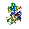
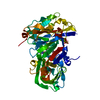
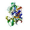
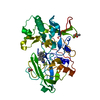
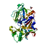
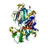
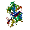
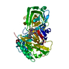
SHEET THE SHEET STRUCTURE OF THIS MOLECULE IS BIFURCATED. IN ORDER TO REPRESENT THIS FEATURE IN ... SHEET THE SHEET STRUCTURE OF THIS MOLECULE IS BIFURCATED. IN ORDER TO REPRESENT THIS FEATURE IN THE SHEET RECORDS BELOW, TWO SHEETS ARE DEFINED.

-
Structure visualization

Structure viewerMolecule:
MolmilJmol/JSmol

Downloads & links

-
Assembly

Deposited unit
A: BETA-SECRETASE 1
hetero molecules


Theoretical massNumber of molelcules
Total (without water)43,6122
Polymers43,1991
Non-polymers4131
Water4,810267
1


  • Idetical with deposited unit
  • defined by author&software
TypeNameSymmetry operationNumber
identity operation1_555x,y,z1
MethodPISA
Unit cell
Length a, b, c (Å)47.990, 75.150, 104.610
Angle α, β, γ (deg.)90.00, 90.00, 90.00
Int Tables number19
Space group name H-MP212121

-
Components

#1: Protein BETA-SECRETASE 1 / ASPARTYL PROTEASE 2 / ASP2 / ASP 2 / BETA-SITE AMYLOID PRECURSOR PROTEIN CLEAVING ENZYME 1 / BETA- ...ASPARTYL PROTEASE 2 / ASP2 / ASP 2 / BETA-SITE AMYLOID PRECURSOR PROTEIN CLEAVING ENZYME 1 / BETA-SITE APP CLEAVING ENZYME 1 / MEMAPSIN-2 / MEMBRANE-ASSOCIATED ASPARTIC PROTEASE 2


Mass: 43198.703 Da / Num. of mol.: 1 / Fragment: RESIDUES 58-445
Source method: isolated from a genetically manipulated source
Source: (gene. exp.) HOMO SAPIENS (human) / Production host: ESCHERICHIA COLI (E. coli) / References: UniProt: P56817, memapsin 2
#2: Chemical ChemComp-2FB / (6R)-6-(4-methoxyphenyl)-2-methyl-6-(3-pyrimidin-5-ylphenyl)pyrrolo[3,4-d][1,3]thiazol-4-amine


Mass: 413.495 Da / Num. of mol.: 1 / Source method: obtained synthetically / Formula: C23H19N5OS
#3: Water ChemComp-HOH / water


Mass: 18.015 Da / Num. of mol.: 267 / Source method: isolated from a natural source / Formula: H2O

-
Experimental details

-
Experiment

ExperimentMethod: X-RAY DIFFRACTION / Number of used crystals: 1

-
Sample preparation

CrystalDensity Matthews: 2.3 Å3/Da / Density % sol: 46 % / Description: NONE
Crystal growpH: 7 / Details: pH 7

-
Data collection

DiffractionMean temperature: 100 K
Diffraction sourceSource: ROTATING ANODE / Wavelength: 1.5418
DetectorType: RIGAKU R-AXIS HTC / Detector: IMAGE PLATE / Date: Dec 13, 2011
RadiationProtocol: SINGLE WAVELENGTH / Monochromatic (M) / Laue (L): M / Scattering type: x-ray
Radiation wavelengthWavelength: 1.5418 Å / Relative weight: 1
ReflectionResolution: 1.6→29.59 Å / Num. obs: 41712 / % possible obs: 84 % / Observed criterion σ(I): 1 / Redundancy: 2.3 % / Biso Wilson estimate: 24.46 Å2 / Rmerge(I) obs: 0.04 / Net I/σ(I): 12.5
Reflection shellResolution: 1.6→1.64 Å / Redundancy: 2 % / Rmerge(I) obs: 0.48 / Mean I/σ(I) obs: 1.7 / % possible all: 74.4

-
Processing

Software
NameVersionClassification
BUSTER2.11.1refinement
MOSFLMdata reduction
SCALAdata scaling
PHASERphasing
RefinementMethod to determine structure: MOLECULAR REPLACEMENT / Resolution: 1.6→29.59 Å / Cor.coef. Fo:Fc: 0.9518 / Cor.coef. Fo:Fc free: 0.9293 / SU R Cruickshank DPI: 0.113 / Cross valid method: THROUGHOUT / σ(F): 0 / SU R Blow DPI: 0.118 / SU Rfree Blow DPI: 0.112 / SU Rfree Cruickshank DPI: 0.11
Details: IDEAL-DIST CONTACT TERM CONTACT SETUP. ALL ATOMS HAVE CCP4 ATOM TYPE FROM LIBRARY
RfactorNum. reflection% reflectionSelection details
Rfree0.229 2128 5.1 %RANDOM
Rwork0.196 ---
obs0.1977 41701 82.26 %-
Displacement parametersBiso mean: 27.47 Å2
Baniso -1Baniso -2Baniso -3
1--1.9157 Å20 Å20 Å2
2---3.8271 Å20 Å2
3---5.7428 Å2
Refine analyzeLuzzati coordinate error obs: 0.205 Å
Refinement stepCycle: LAST / Resolution: 1.6→29.59 Å
ProteinNucleic acidLigandSolventTotal
Num. atoms2956 0 30 267 3253
Refine LS restraints
Refine-IDTypeDev idealNumberRestraint functionWeight
X-RAY DIFFRACTIONt_bond_d0.013112HARMONIC2
X-RAY DIFFRACTIONt_angle_deg1.134248HARMONIC2
X-RAY DIFFRACTIONt_dihedral_angle_d1040SINUSOIDAL2
X-RAY DIFFRACTIONt_incorr_chiral_ct
X-RAY DIFFRACTIONt_pseud_angle
X-RAY DIFFRACTIONt_trig_c_planes72HARMONIC2
X-RAY DIFFRACTIONt_gen_planes462HARMONIC5
X-RAY DIFFRACTIONt_it3112HARMONIC20
X-RAY DIFFRACTIONt_nbd
X-RAY DIFFRACTIONt_omega_torsion4.14
X-RAY DIFFRACTIONt_other_torsion15.41
X-RAY DIFFRACTIONt_improper_torsion
X-RAY DIFFRACTIONt_chiral_improper_torsion398SEMIHARMONIC5
X-RAY DIFFRACTIONt_sum_occupancies
X-RAY DIFFRACTIONt_utility_distance
X-RAY DIFFRACTIONt_utility_angle
X-RAY DIFFRACTIONt_utility_torsion
X-RAY DIFFRACTIONt_ideal_dist_contact3729SEMIHARMONIC4
LS refinement shellResolution: 1.6→1.64 Å / Total num. of bins used: 20
RfactorNum. reflection% reflection
Rfree0.3975 130 4.85 %
Rwork0.3722 2548 -
all0.3734 2678 -
obs--82.26 %

+
About Yorodumi

-
News

-
Feb 9, 2022. New format data for meta-information of EMDB entries

New format data for meta-information of EMDB entries

  • Version 3 of the EMDB header file is now the official format.
  • The previous official version 1.9 will be removed from the archive.

Related info.:EMDB header

External links:wwPDB to switch to version 3 of the EMDB data model

-
Aug 12, 2020. Covid-19 info

Covid-19 info

URL: https://pdbjlvh1.pdbj.org/emnavi/covid19.php

New page: Covid-19 featured information page in EM Navigator.

Related info.:Covid-19 info / Mar 5, 2020. Novel coronavirus structure data

+
Mar 5, 2020. Novel coronavirus structure data

Novel coronavirus structure data

Related info.:Yorodumi Speices / Aug 12, 2020. Covid-19 info

External links:COVID-19 featured content - PDBj / Molecule of the Month (242):Coronavirus Proteases

+
Jan 31, 2019. EMDB accession codes are about to change! (news from PDBe EMDB page)

EMDB accession codes are about to change! (news from PDBe EMDB page)

  • The allocation of 4 digits for EMDB accession codes will soon come to an end. Whilst these codes will remain in use, new EMDB accession codes will include an additional digit and will expand incrementally as the available range of codes is exhausted. The current 4-digit format prefixed with “EMD-” (i.e. EMD-XXXX) will advance to a 5-digit format (i.e. EMD-XXXXX), and so on. It is currently estimated that the 4-digit codes will be depleted around Spring 2019, at which point the 5-digit format will come into force.
  • The EM Navigator/Yorodumi systems omit the EMD- prefix.

Related info.:Q: What is EMD? / ID/Accession-code notation in Yorodumi/EM Navigator

External links:EMDB Accession Codes are Changing Soon! / Contact to PDBj

+
Jul 12, 2017. Major update of PDB

Major update of PDB

  • wwPDB released updated PDB data conforming to the new PDBx/mmCIF dictionary.
  • This is a major update changing the version number from 4 to 5, and with Remediation, in which all the entries are updated.
  • In this update, many items about electron microscopy experimental information are reorganized (e.g. em_software).
  • Now, EM Navigator and Yorodumi are based on the updated data.

External links:wwPDB Remediation / Enriched Model Files Conforming to OneDep Data Standards Now Available in the PDB FTP Archive

-
Yorodumi

Thousand views of thousand structures

  • Yorodumi is a browser for structure data from EMDB, PDB, SASBDB, etc.
  • This page is also the successor to EM Navigator detail page, and also detail information page/front-end page for Omokage search.
  • The word "yorodu" (or yorozu) is an old Japanese word meaning "ten thousand". "mi" (miru) is to see.

Related info.:EMDB / PDB / SASBDB / Comparison of 3 databanks / Yorodumi Search / Aug 31, 2016. New EM Navigator & Yorodumi / Yorodumi Papers / Jmol/JSmol / Function and homology information / Changes in new EM Navigator and Yorodumi

Read more