 Movie
Movie Controller

[English] 日本語
 Yorodumi
- PDB-1ujj: VHS domain of human GGA1 complexed with C-terminal peptide from BACE -
+ Open data

- Basic information
| Entry | Database: PDB / ID: 1ujj | ||||||
|---|---|---|---|---|---|---|---|
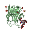
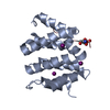
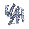
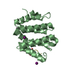
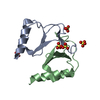
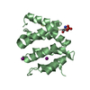
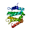
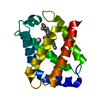
| Title | VHS domain of human GGA1 complexed with C-terminal peptide from BACE | ||||||
 Components Components |
| ||||||
 Keywords Keywords | PROTEIN TRANSPORT/Hydrolase / PROTEIN-PEPTIDE COMPLEX / PROTEIN TRANSPORT / ADAPTOR PROTEIN / PROTEIN TRANSPORT-Hydrolase COMPLEX | ||||||
| Function / homology |  Function and homology information Function and homology informationprotein localization to ciliary membrane / Golgi to plasma membrane transport / Golgi to plasma membrane protein transport / TBC/RABGAPs / protein localization to cell surface / retrograde transport, endosome to Golgi / memapsin 2 / Golgi-associated vesicle lumen / beta-aspartyl-peptidase activity / signaling receptor ligand precursor processing ...protein localization to ciliary membrane / Golgi to plasma membrane transport / Golgi to plasma membrane protein transport / TBC/RABGAPs / protein localization to cell surface / retrograde transport, endosome to Golgi / memapsin 2 / Golgi-associated vesicle lumen / beta-aspartyl-peptidase activity / signaling receptor ligand precursor processing / amyloid-beta formation / amyloid precursor protein catabolic process / membrane protein ectodomain proteolysis / amyloid-beta metabolic process / detection of mechanical stimulus involved in sensory perception of pain / response to insulin-like growth factor stimulus / prepulse inhibition / cellular response to manganese ion / multivesicular body / swimming behavior / phosphatidylinositol binding / cellular response to copper ion / presynaptic modulation of chemical synaptic transmission / hippocampal mossy fiber to CA3 synapse / protein catabolic process / protein serine/threonine kinase binding / ubiquitin binding / trans-Golgi network / protein processing / intracellular protein transport / recycling endosome / response to lead ion / small GTPase binding / cellular response to amyloid-beta / positive regulation of protein catabolic process / synaptic vesicle / intracellular protein localization / late endosome / peptidase activity / positive regulation of neuron apoptotic process / amyloid-beta binding / early endosome membrane / endopeptidase activity / amyloid fibril formation / aspartic-type endopeptidase activity / early endosome / lysosome / endosome / endosome membrane / membrane raft / endoplasmic reticulum lumen / Amyloid fiber formation / axon / neuronal cell body / dendrite / enzyme binding / cell surface / Golgi apparatus / protein-containing complex / proteolysis / nucleoplasm / membrane / plasma membrane / cytosol Similarity search - Function | ||||||
| Biological species |  Homo sapiens (human) Homo sapiens (human) | ||||||
| Method |  X-RAY DIFFRACTION / SYNCHROTRON / MOLECULAR REPLACEMENT / Resolution: 2.6 Å X-RAY DIFFRACTION / SYNCHROTRON / MOLECULAR REPLACEMENT / Resolution: 2.6 Å | ||||||
 Authors Authors | Shiba, T. / Kametaka, S. / Kawasaki, M. / Shibata, M. / Waguri, S. / Uchiyama, Y. / Wakatsuki, S. | ||||||
 Citation Citation | Journal: TRAFFIC / Year: 2004 Title: Insights into the Phosphoregulation of beta-Secretase Sorting Signal by the VHS Domain of GGA1 Authors: Shiba, T. / Kametaka, S. / Kawasaki, M. / Shibata, M. / Waguri, S. / Uchiyama, Y. / Wakatsuki, S. | ||||||
| History |
|
- Structure visualization
| Structure viewer | Molecule: MolmilJmol/JSmol |
|---|
- Downloads & links
-Download
| PDBx/mmCIF format | 1ujj.cif.gz | 66 KB | Display | PDBx/mmCIF format |
|---|---|---|---|---|
| PDB format | pdb1ujj.ent.gz | 50.6 KB | Display | PDB format |
| PDBx/mmJSON format | 1ujj.json.gz | Tree view | PDBx/mmJSON format | |
| Others |  Other downloads Other downloads |
-Validation report
| Arichive directory | https://data.pdbj.org/pub/pdb/validation_reports/uj/1ujjftp://data.pdbj.org/pub/pdb/validation_reports/uj/1ujj | HTTPS FTP |
|---|
-Related structure data
| Related structure data | 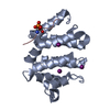 1ujkC 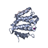 1jwgS S: Starting model for refinement C: citing same article ( |
|---|---|
| Similar structure data |


-Links
 PDBj
PDBj


- Assembly
| Deposited unit | 
| ||||||||
|---|---|---|---|---|---|---|---|---|---|
| 1 | 
| ||||||||
| 2 | 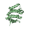
| ||||||||
| Unit cell |
|
-Components
| #1: Protein | Mass: 16814.436 Da / Num. of mol.: 2 / Fragment: VHS domain, N-terminal domain Source method: isolated from a genetically manipulated source Source: (gene. exp.) Homo sapiens (human) / Plasmid: pGEX4T-2 / Species (production host): Escherichia coli / Production host:  #2: Protein/peptide | | Mass: 1390.496 Da / Num. of mol.: 1 / Source method: obtained synthetically / Details: Synthesized peptide References: UniProt: P56817, Hydrolases; Acting on peptide bonds (peptidases); Aspartic endopeptidases Has protein modification | Y | |
|---|
-Experimental details
-Experiment
| Experiment | Method: X-RAY DIFFRACTION / Number of used crystals: 1 |
|---|
- Sample preparation
| Crystal | Density Matthews: 2.74 Å3/Da / Density % sol: 54.69 % |
|---|---|
| Crystal grow | Temperature: 293 K / Method: vapor diffusion, hanging drop / pH: 8.6 Details: PEG 5000MME, di-Ammonium hydrogen phosphate, Tris-HCl, pH 8.6, VAPOR DIFFUSION, HANGING DROP, temperature 293K |
-Data collection
| Diffraction | Mean temperature: 100 K |
|---|---|
| Diffraction source | Source: SYNCHROTRON / Site: Photon Factory  / Beamline: AR-NW12A / Wavelength: 1 Å / Beamline: AR-NW12A / Wavelength: 1 Å |
| Detector | Type: ADSC QUANTUM 210 / Detector: CCD / Date: Jun 14, 2003 |
| Radiation | Monochromator: Si(111) / Protocol: SINGLE WAVELENGTH / Monochromatic (M) / Laue (L): M / Scattering type: x-ray |
| Radiation wavelength | Wavelength: 1 Å / Relative weight: 1 |
| Reflection | Resolution: 2.6→40 Å / Num. all: 10872 / Num. obs: 10857 / % possible obs: 99.4 % / Rmerge(I) obs: 0.059 / Net I/σ(I): 20.7 |
| Reflection shell | Resolution: 2.6→2.69 Å / % possible all: 99.7 |
- Processing
| Software |
| |||||||||||||||||||||||||
|---|---|---|---|---|---|---|---|---|---|---|---|---|---|---|---|---|---|---|---|---|---|---|---|---|---|---|
| Refinement | Method to determine structure: MOLECULAR REPLACEMENT Starting model: PDB ENTRY 1JWG Resolution: 2.6→40 Å / Cross valid method: THROUGHOUT / σ(F): 0 / Stereochemistry target values: Engh & Huber
| |||||||||||||||||||||||||
| Refinement step | Cycle: LAST / Resolution: 2.6→40 Å
| |||||||||||||||||||||||||
| Refine LS restraints |
|
