 Movie
Movie Controller

[English] 日本語
 Yorodumi
- PDB-1jwg: VHS Domain of human GGA1 complexed with cation-independent M6PR C... -
+ Open data

- Basic information
| Entry | Database: PDB / ID: 1jwg | ||||||
|---|---|---|---|---|---|---|---|
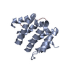
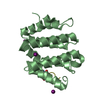
| Title | VHS Domain of human GGA1 complexed with cation-independent M6PR C-terminal Peptide | ||||||
 Components Components |
| ||||||
 Keywords Keywords | PROTEIN TRANSPORT/PROTEIN BINDING / SUPER HELIX / PROTEIN-PEPTIDE COMPLEX / PROTEIN TRANSPORT-PROTEIN BINDING COMPLEX | ||||||
| Function / homology |  Function and homology information Function and homology informationprotein localization to ciliary membrane / Retrograde transport at the Trans-Golgi-Network / insulin-like growth factor receptor activity / clathrin coat / response to tetrachloromethane / retromer complex binding / insulin-like growth factor binding / insulin-like growth factor II binding / Golgi to plasma membrane transport / trans-Golgi network transport vesicle ...protein localization to ciliary membrane / Retrograde transport at the Trans-Golgi-Network / insulin-like growth factor receptor activity / clathrin coat / response to tetrachloromethane / retromer complex binding / insulin-like growth factor binding / insulin-like growth factor II binding / Golgi to plasma membrane transport / trans-Golgi network transport vesicle / host-mediated activation of viral process / protein targeting to lysosome / retinoic acid binding / Golgi to plasma membrane protein transport / TBC/RABGAPs / retrograde transport, endosome to Golgi / protein localization to cell surface / nuclear envelope lumen / Golgi Associated Vesicle Biogenesis / D-mannose binding / animal organ regeneration / response to retinoic acid / endocytic vesicle / G-protein alpha-subunit binding / transport vesicle / phosphatidylinositol binding / post-embryonic development / receptor-mediated endocytosis / secretory granule membrane / liver development / trans-Golgi network membrane / ubiquitin binding / trans-Golgi network / clathrin-coated endocytic vesicle membrane / phosphoprotein binding / intracellular protein transport / small GTPase binding / intracellular protein localization / late endosome / Cargo recognition for clathrin-mediated endocytosis / Clathrin-mediated endocytosis / signaling receptor activity / early endosome membrane / spermatogenesis / early endosome / endosome / endosome membrane / positive regulation of apoptotic process / G protein-coupled receptor signaling pathway / Amyloid fiber formation / Golgi membrane / focal adhesion / Neutrophil degranulation / perinuclear region of cytoplasm / Golgi apparatus / enzyme binding / cell surface / signal transduction / protein-containing complex / extracellular exosome / membrane / identical protein binding / plasma membrane / cytosol Similarity search - Function | ||||||
| Biological species |  Homo sapiens (human) Homo sapiens (human) | ||||||
| Method |  X-RAY DIFFRACTION / SYNCHROTRON / MOLECULAR REPLACEMENT / Resolution: 2 Å X-RAY DIFFRACTION / SYNCHROTRON / MOLECULAR REPLACEMENT / Resolution: 2 Å | ||||||
 Authors Authors | Shiba, T. / Takatsu, H. / Nogi, T. / Matsugaki, N. / Kawasaki, M. / Igarashi, N. / Suzuki, M. / Kato, R. / Earnest, T. / Nakayama, K. / Wakatsuki, S. | ||||||
 Citation Citation | Journal: Nature / Year: 2002 Title: Structural basis for recognition of acidic-cluster dileucine sequence by GGA1. Authors: Shiba, T. / Takatsu, H. / Nogi, T. / Matsugaki, N. / Kawasaki, M. / Igarashi, N. / Suzuki, M. / Kato, R. / Earnest, T. / Nakayama, K. / Wakatsuki, S. | ||||||
| History |
|
- Structure visualization
| Structure viewer | Molecule: MolmilJmol/JSmol |
|---|
- Downloads & links
-Download
| PDBx/mmCIF format | 1jwg.cif.gz | 75 KB | Display | PDBx/mmCIF format |
|---|---|---|---|---|
| PDB format | pdb1jwg.ent.gz | 56.8 KB | Display | PDB format |
| PDBx/mmJSON format | 1jwg.json.gz | Tree view | PDBx/mmJSON format | |
| Others |  Other downloads Other downloads |
-Validation report
| Arichive directory | https://data.pdbj.org/pub/pdb/validation_reports/jw/1jwgftp://data.pdbj.org/pub/pdb/validation_reports/jw/1jwg | HTTPS FTP |
|---|
-Related structure data
| Related structure data | 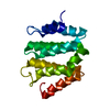 1jwfSC S: Starting model for refinement C: citing same article ( |
|---|---|
| Similar structure data |


-Links
 PDBj
PDBj


- Assembly
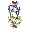
| Deposited unit | 
| ||||||||
|---|---|---|---|---|---|---|---|---|---|
| 1 | 
| ||||||||
| 2 | 
| ||||||||
| Unit cell |
|
-Components
| #1: Protein | Mass: 16814.436 Da / Num. of mol.: 2 / Fragment: VHS DOMAIN(N-terminal domain) Source method: isolated from a genetically manipulated source Source: (gene. exp.) Homo sapiens (human) / Plasmid: pgex4t-2 / Production host:  #2: Protein/peptide | Mass: 1544.577 Da / Num. of mol.: 2 / Fragment: C-terminal fragment / Source method: obtained synthetically / Details: This sequence occurs naturally in humans. / References: UniProt: P11717 #3: Chemical | ChemComp-IOD /   Mass: 126.904 Da / Num. of mol.: 6 / Source method: obtained synthetically / Formula: I Mass: 126.904 Da / Num. of mol.: 6 / Source method: obtained synthetically / Formula: I#4: Water | ChemComp-HOH / |  Mass: 18.015 Da / Num. of mol.: 198 / Source method: isolated from a natural source / Formula: H2O Mass: 18.015 Da / Num. of mol.: 198 / Source method: isolated from a natural source / Formula: H2OHas protein modification | Y | |
|---|
-Experimental details
-Experiment
| Experiment | Method: X-RAY DIFFRACTION / Number of used crystals: 1 |
|---|
- Sample preparation
| Crystal | Density Matthews: 2.55 Å3/Da / Density % sol: 51.74 % | ||||||||||||||||||||||||||||||||||||||||||
|---|---|---|---|---|---|---|---|---|---|---|---|---|---|---|---|---|---|---|---|---|---|---|---|---|---|---|---|---|---|---|---|---|---|---|---|---|---|---|---|---|---|---|---|
| Crystal grow | Temperature: 293 K / Method: vapor diffusion, hanging drop / pH: 7.5 Details: PEG 3350, ammonium iodide, Tris, pH 7.5, VAPOR DIFFUSION, HANGING DROP, temperature 293K | ||||||||||||||||||||||||||||||||||||||||||
| Crystal grow | *PLUS pH: 8 | ||||||||||||||||||||||||||||||||||||||||||
| Components of the solutions | *PLUS
|
-Data collection
| Diffraction | Mean temperature: 100 K |
|---|---|
| Diffraction source | Source: SYNCHROTRON / Site: ALS  / Beamline: 5.0.2 / Wavelength: 1 Å / Beamline: 5.0.2 / Wavelength: 1 Å |
| Detector | Type: ADSC QUANTUM 4 / Detector: CCD / Date: Aug 16, 2001 |
| Radiation | Monochromator: Si(111) / Protocol: SINGLE WAVELENGTH / Monochromatic (M) / Laue (L): M / Scattering type: x-ray |
| Radiation wavelength | Wavelength: 1 Å / Relative weight: 1 |
| Reflection | Resolution: 2→30 Å / Num. all: 24632 / Num. obs: 24632 / % possible obs: 99.6 % |
| Reflection shell | Resolution: 2→2.11 Å / % possible all: 99.3 |
| Reflection | *PLUS Num. obs: 25975 / Num. measured all: 167862 / Rmerge(I) obs: 0.067 |
- Processing
| Software |
| ||||||||||||||||||||||||||||||||||||||||||||||||||||||||||||||||||||||||||||||||||||||||||||||||||||||||||||||||||||||||||||||||||
|---|---|---|---|---|---|---|---|---|---|---|---|---|---|---|---|---|---|---|---|---|---|---|---|---|---|---|---|---|---|---|---|---|---|---|---|---|---|---|---|---|---|---|---|---|---|---|---|---|---|---|---|---|---|---|---|---|---|---|---|---|---|---|---|---|---|---|---|---|---|---|---|---|---|---|---|---|---|---|---|---|---|---|---|---|---|---|---|---|---|---|---|---|---|---|---|---|---|---|---|---|---|---|---|---|---|---|---|---|---|---|---|---|---|---|---|---|---|---|---|---|---|---|---|---|---|---|---|---|---|---|---|
| Refinement | Method to determine structure: MOLECULAR REPLACEMENT Starting model: PDB ENTRY 1JWF Resolution: 2→30 Å / Cor.coef. Fo:Fc: 0.933 / SU B: 4.254 / SU ML: 0.121 / Cross valid method: THROUGHOUT / σ(F): 0 / ESU R: 0.194 / ESU R Free: 0.171 / Stereochemistry target values: MAXIMUM LIKELIHOOD
| ||||||||||||||||||||||||||||||||||||||||||||||||||||||||||||||||||||||||||||||||||||||||||||||||||||||||||||||||||||||||||||||||||
| Solvent computation | Ion probe radii: 0.8 Å / Shrinkage radii: 0.8 Å / VDW probe radii: 1.4 Å / Solvent model: BABINET MODEL WITH MASK | ||||||||||||||||||||||||||||||||||||||||||||||||||||||||||||||||||||||||||||||||||||||||||||||||||||||||||||||||||||||||||||||||||
| Displacement parameters | Biso mean: 39.099 Å2
| ||||||||||||||||||||||||||||||||||||||||||||||||||||||||||||||||||||||||||||||||||||||||||||||||||||||||||||||||||||||||||||||||||
| Refinement step | Cycle: LAST / Resolution: 2→30 Å
| ||||||||||||||||||||||||||||||||||||||||||||||||||||||||||||||||||||||||||||||||||||||||||||||||||||||||||||||||||||||||||||||||||
| Refine LS restraints |
| ||||||||||||||||||||||||||||||||||||||||||||||||||||||||||||||||||||||||||||||||||||||||||||||||||||||||||||||||||||||||||||||||||
| LS refinement shell | Resolution: 2→2.052 Å / Total num. of bins used: 20
| ||||||||||||||||||||||||||||||||||||||||||||||||||||||||||||||||||||||||||||||||||||||||||||||||||||||||||||||||||||||||||||||||||
| Refinement | *PLUS Highest resolution: 2 Å / σ(F): 0 / % reflection Rfree: 5 % | ||||||||||||||||||||||||||||||||||||||||||||||||||||||||||||||||||||||||||||||||||||||||||||||||||||||||||||||||||||||||||||||||||
| Solvent computation | *PLUS | ||||||||||||||||||||||||||||||||||||||||||||||||||||||||||||||||||||||||||||||||||||||||||||||||||||||||||||||||||||||||||||||||||
| Displacement parameters | *PLUS | ||||||||||||||||||||||||||||||||||||||||||||||||||||||||||||||||||||||||||||||||||||||||||||||||||||||||||||||||||||||||||||||||||
| LS refinement shell | *PLUS Highest resolution: 2 Å / Lowest resolution: 2.05 Å / Rfactor Rfree: 0.272 / Rfactor Rwork: 0.234 |
