 Movie
Movie Controller

[English] 日本語
 Yorodumi
- PDB-3qbh: Structure based design, synthesis and SAR of cyclic hydroxyethyla... -
+ Open data

- Basic information
| Entry | Database: PDB / ID: 3qbh | ||||||
|---|---|---|---|---|---|---|---|
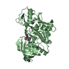
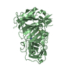
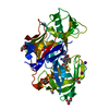
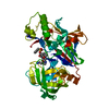
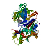
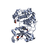
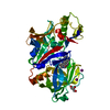
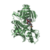
| Title | Structure based design, synthesis and SAR of cyclic hydroxyethylamine (HEA) BACE-1 inhibitors | ||||||
 Components Components | Beta-secretase 1 | ||||||
 Keywords Keywords | HYDROLASE/HYDROLASE Inhibitor / Enzyme inhibitor complex / HYDROLASE / HYDROLASE-HYDROLASE Inhibitor complex | ||||||
| Function / homology |  Function and homology information Function and homology informationmemapsin 2 / Golgi-associated vesicle lumen / beta-aspartyl-peptidase activity / signaling receptor ligand precursor processing / amyloid-beta formation / amyloid precursor protein catabolic process / membrane protein ectodomain proteolysis / amyloid-beta metabolic process / detection of mechanical stimulus involved in sensory perception of pain / response to insulin-like growth factor stimulus ...memapsin 2 / Golgi-associated vesicle lumen / beta-aspartyl-peptidase activity / signaling receptor ligand precursor processing / amyloid-beta formation / amyloid precursor protein catabolic process / membrane protein ectodomain proteolysis / amyloid-beta metabolic process / detection of mechanical stimulus involved in sensory perception of pain / response to insulin-like growth factor stimulus / prepulse inhibition / swimming behavior / cellular response to manganese ion / multivesicular body / cellular response to copper ion / presynaptic modulation of chemical synaptic transmission / hippocampal mossy fiber to CA3 synapse / protein serine/threonine kinase binding / trans-Golgi network / protein processing / recycling endosome / response to lead ion / cellular response to amyloid-beta / late endosome / peptidase activity / positive regulation of neuron apoptotic process / synaptic vesicle / amyloid-beta binding / endopeptidase activity / aspartic-type endopeptidase activity / amyloid fibril formation / early endosome / lysosome / endosome / endosome membrane / membrane raft / endoplasmic reticulum lumen / Amyloid fiber formation / axon / neuronal cell body / dendrite / Golgi apparatus / enzyme binding / cell surface / proteolysis / membrane / plasma membrane Similarity search - Function | ||||||
| Biological species |  Homo sapiens (human) Homo sapiens (human) | ||||||
| Method |  X-RAY DIFFRACTION / SYNCHROTRON / FOURIER SYNTHESIS / Resolution: 2.24 Å X-RAY DIFFRACTION / SYNCHROTRON / FOURIER SYNTHESIS / Resolution: 2.24 Å | ||||||
 Authors Authors | Rondeau, J.M. | ||||||
 Citation Citation | Journal: Bioorg.Med.Chem.Lett. / Year: 2011 Title: Structure based design, synthesis and SAR of cyclic hydroxyethylamine (HEA) BACE-1 inhibitors. Authors: Rueeger, H. / Rondeau, J.M. / McCarthy, C. / Mobitz, H. / Tintelnot-Blomley, M. / Neumann, U. / Desrayaud, S. #1: Journal: Bioorg.Med.Chem.Lett. / Year: 2009Title: Structure-Based Design of Macrocyclic Peptidomimetic beta-Secretase (BACE-1) Inhibitors Authors: Machauer, R. / Veenstra, S. / Rondeau, J.M. / Tintelnot-Blomley, M. / Betschart, C. / Neumann, U. / Paganetti, P. #2: Journal: Bioorg.Med.Chem.Lett. / Year: 2009Title: Macrocyclic peptidomimetic beta-secretase (BACE-1) inhibitors with activity in vivo Authors: Machauer, R. / Laumen, K. / Veenstra, S. / Rondeau, J.M. / Tintelnot-Blomley, M. / Betschart, C. / Jaton, A.-L. / Desrayaud, S. / Staufenbiel, M. / Rabe, S. / Paganetti, P. / Neumann, U. #3: Journal: Bioorg.Med.Chem.Lett. / Year: 2010Title: Macrocyclic BACE-1 inhibitors acutely reduce Abeta in brain after po application Authors: Lerchner, A. / Machauer, R. / Betschart, C. / Veenstra, S. / Rueeger, H. / McCarthy, C. / Tintelnot-Blomley, M. / Jaton, A.-L. / Rabe, S. / Desrayaud, S. / Enz, A. / Staufenbiel, M. / ...Authors: Lerchner, A. / Machauer, R. / Betschart, C. / Veenstra, S. / Rueeger, H. / McCarthy, C. / Tintelnot-Blomley, M. / Jaton, A.-L. / Rabe, S. / Desrayaud, S. / Enz, A. / Staufenbiel, M. / Paganetti, P. / Rondeau, J.M. / Neumann, U. | ||||||
| History |
|
- Structure visualization
| Structure viewer | Molecule: MolmilJmol/JSmol |
|---|
- Downloads & links
-Download
| PDBx/mmCIF format | 3qbh.cif.gz | 245.6 KB | Display | PDBx/mmCIF format |
|---|---|---|---|---|
| PDB format | pdb3qbh.ent.gz | 196.4 KB | Display | PDB format |
| PDBx/mmJSON format | 3qbh.json.gz | Tree view | PDBx/mmJSON format | |
| Others |  Other downloads Other downloads |
-Validation report
| Arichive directory | https://data.pdbj.org/pub/pdb/validation_reports/qb/3qbhftp://data.pdbj.org/pub/pdb/validation_reports/qb/3qbh | HTTPS FTP |
|---|
-Related structure data


-Links
 PDBj
PDBj

- Assembly
| Deposited unit | 
| ||||||||
|---|---|---|---|---|---|---|---|---|---|
| 1 | 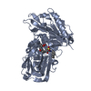
| ||||||||
| 2 | 
| ||||||||
| 3 | 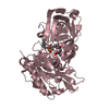
| ||||||||
| Unit cell |
|
-Components
| #1: Protein | Mass: 44777.336 Da / Num. of mol.: 3 / Fragment: UNP residues 48-447 Source method: isolated from a genetically manipulated source Details: Refolded / Source: (gene. exp.) Homo sapiens (human) / Gene: BACE, BACE1, KIAA1149 / Plasmid: pET24 / Production host:  #2: Chemical |   Mass: 544.703 Da / Num. of mol.: 3 / Source method: obtained synthetically / Formula: C29H40N2O6S Mass: 544.703 Da / Num. of mol.: 3 / Source method: obtained synthetically / Formula: C29H40N2O6S#3: Water | ChemComp-HOH / |  Mass: 18.015 Da / Num. of mol.: 501 / Source method: isolated from a natural source / Formula: H2O Mass: 18.015 Da / Num. of mol.: 501 / Source method: isolated from a natural source / Formula: H2OHas protein modification | Y | |
|---|
-Experimental details
-Experiment
| Experiment | Method: X-RAY DIFFRACTION / Number of used crystals: 1 |
|---|
- Sample preparation
| Crystal | Density Matthews: 3.08 Å3/Da / Density % sol: 60.07 % |
|---|---|
| Crystal grow | Temperature: 292 K / Method: vapor diffusion, hanging drop / pH: 5 Details: 1.0M Ammonium phosphate, 0.1M sodium citrate pH 5.0, VAPOR DIFFUSION, HANGING DROP, temperature 292K |
-Data collection
| Diffraction | Mean temperature: 100 K | ||||||||||||||||||||||||||||||||||||||||||||||||||||||||||||||||||||||||||||||||||||||||||||||||||||||||||||||||||||||||||||||||||||||||||||||||||||||||||||||||
|---|---|---|---|---|---|---|---|---|---|---|---|---|---|---|---|---|---|---|---|---|---|---|---|---|---|---|---|---|---|---|---|---|---|---|---|---|---|---|---|---|---|---|---|---|---|---|---|---|---|---|---|---|---|---|---|---|---|---|---|---|---|---|---|---|---|---|---|---|---|---|---|---|---|---|---|---|---|---|---|---|---|---|---|---|---|---|---|---|---|---|---|---|---|---|---|---|---|---|---|---|---|---|---|---|---|---|---|---|---|---|---|---|---|---|---|---|---|---|---|---|---|---|---|---|---|---|---|---|---|---|---|---|---|---|---|---|---|---|---|---|---|---|---|---|---|---|---|---|---|---|---|---|---|---|---|---|---|---|---|---|---|
| Diffraction source | Source: SYNCHROTRON / Site: SLS  / Beamline: X10SA / Wavelength: 0.97629 Å / Beamline: X10SA / Wavelength: 0.97629 Å | ||||||||||||||||||||||||||||||||||||||||||||||||||||||||||||||||||||||||||||||||||||||||||||||||||||||||||||||||||||||||||||||||||||||||||||||||||||||||||||||||
| Detector | Type: MARMOSAIC 225 mm CCD / Detector: CCD / Date: Apr 21, 2005 | ||||||||||||||||||||||||||||||||||||||||||||||||||||||||||||||||||||||||||||||||||||||||||||||||||||||||||||||||||||||||||||||||||||||||||||||||||||||||||||||||
| Radiation | Monochromator: SAGITALLY FOCUSED Si(111) / Protocol: SINGLE WAVELENGTH / Monochromatic (M) / Laue (L): M / Scattering type: x-ray | ||||||||||||||||||||||||||||||||||||||||||||||||||||||||||||||||||||||||||||||||||||||||||||||||||||||||||||||||||||||||||||||||||||||||||||||||||||||||||||||||
| Radiation wavelength | Wavelength: 0.97629 Å / Relative weight: 1 | ||||||||||||||||||||||||||||||||||||||||||||||||||||||||||||||||||||||||||||||||||||||||||||||||||||||||||||||||||||||||||||||||||||||||||||||||||||||||||||||||
| Reflection | Resolution: 2.24→80 Å / Num. all: 78093 / Num. obs: 78093 / % possible obs: 99.6 % / Observed criterion σ(F): 0 / Observed criterion σ(I): -3 / Redundancy: 7.6 % / Biso Wilson estimate: 48.891 Å2 / Rmerge(I) obs: 0.063 / Net I/σ(I): 18.6 | ||||||||||||||||||||||||||||||||||||||||||||||||||||||||||||||||||||||||||||||||||||||||||||||||||||||||||||||||||||||||||||||||||||||||||||||||||||||||||||||||
| Reflection shell | Diffraction-ID: 1
|
- Processing
| Software |
| ||||||||||||||||||||||||||||||||||||
|---|---|---|---|---|---|---|---|---|---|---|---|---|---|---|---|---|---|---|---|---|---|---|---|---|---|---|---|---|---|---|---|---|---|---|---|---|---|
| Refinement | Method to determine structure: FOURIER SYNTHESIS / Resolution: 2.24→80 Å / Rfactor Rfree error: 0.002 / Occupancy max: 1 / Occupancy min: 1 / FOM work R set: 0.8558 / Data cutoff high absF: 2328977 / Data cutoff low absF: 0 / Isotropic thermal model: RESTRAINED / Cross valid method: THROUGHOUT / σ(F): 0 / Stereochemistry target values: Engh & Huber
| ||||||||||||||||||||||||||||||||||||
| Solvent computation | Solvent model: FLAT MODEL / Bsol: 51.9376 Å2 / ksol: 0.3693 e/Å3 | ||||||||||||||||||||||||||||||||||||
| Displacement parameters | Biso max: 116.77 Å2 / Biso mean: 48.5611 Å2 / Biso min: 22.7 Å2
| ||||||||||||||||||||||||||||||||||||
| Refine analyze |
| ||||||||||||||||||||||||||||||||||||
| Refinement step | Cycle: LAST / Resolution: 2.24→80 Å
| ||||||||||||||||||||||||||||||||||||
| Refine LS restraints |
| ||||||||||||||||||||||||||||||||||||
| LS refinement shell | Resolution: 2.24→2.38 Å / Rfactor Rfree error: 0.002 / Total num. of bins used: 6
| ||||||||||||||||||||||||||||||||||||
| Xplor file |
|
