 Movie
Movie Controller

[English] 日本語
 Yorodumi
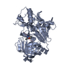
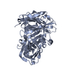
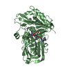
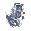
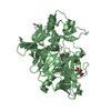
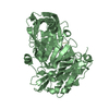
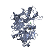
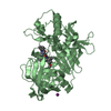
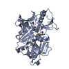
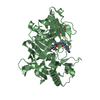
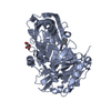
- PDB-1xn3: Crystal structure of Beta-secretase bound to a long inhibitor wit... -
+ Open data

- Basic information
| Entry | Database: PDB / ID: 1xn3 | ||||||
|---|---|---|---|---|---|---|---|
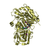
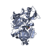
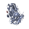
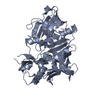
| Title | Crystal structure of Beta-secretase bound to a long inhibitor with additional upstream residues. | ||||||
 Components Components |
| ||||||
 Keywords Keywords | HYDROLASE/HYDROLASE INHIBITOR / BACE / Alzheimer's disease / HYDROLASE-HYDROLASE INHIBITOR complex | ||||||
| Function / homology |  Function and homology information Function and homology informationmemapsin 2 / Golgi-associated vesicle lumen / beta-aspartyl-peptidase activity / signaling receptor ligand precursor processing / amyloid-beta formation / amyloid precursor protein catabolic process / membrane protein ectodomain proteolysis / amyloid-beta metabolic process / detection of mechanical stimulus involved in sensory perception of pain / response to insulin-like growth factor stimulus ...memapsin 2 / Golgi-associated vesicle lumen / beta-aspartyl-peptidase activity / signaling receptor ligand precursor processing / amyloid-beta formation / amyloid precursor protein catabolic process / membrane protein ectodomain proteolysis / amyloid-beta metabolic process / detection of mechanical stimulus involved in sensory perception of pain / response to insulin-like growth factor stimulus / prepulse inhibition / swimming behavior / cellular response to manganese ion / multivesicular body / cellular response to copper ion / presynaptic modulation of chemical synaptic transmission / hippocampal mossy fiber to CA3 synapse / protein serine/threonine kinase binding / trans-Golgi network / protein processing / recycling endosome / response to lead ion / cellular response to amyloid-beta / late endosome / peptidase activity / positive regulation of neuron apoptotic process / synaptic vesicle / amyloid-beta binding / endopeptidase activity / aspartic-type endopeptidase activity / amyloid fibril formation / early endosome / lysosome / endosome / endosome membrane / membrane raft / endoplasmic reticulum lumen / Amyloid fiber formation / axon / neuronal cell body / dendrite / Golgi apparatus / enzyme binding / cell surface / proteolysis / membrane / plasma membrane Similarity search - Function | ||||||
| Biological species |  Homo sapiens (human) Homo sapiens (human) | ||||||
| Method |  X-RAY DIFFRACTION / MOLECULAR REPLACEMENT / Resolution: 2 Å X-RAY DIFFRACTION / MOLECULAR REPLACEMENT / Resolution: 2 Å | ||||||
 Authors Authors | Turner III, R.T. / Hong, L. / Koelsch, G. / Ghosh, A.K. / Tang, J. | ||||||
 Citation Citation | Journal: Biochemistry / Year: 2005 Title: Structural locations and functional roles of new subsites S5, S6, and S7 in memapsin 2 (beta-secretase). Authors: Turner III, R.T. / Hong, L. / Koelsch, G. / Ghosh, A.K. / Tang, J. | ||||||
| History |
|
- Structure visualization
| Structure viewer | Molecule: MolmilJmol/JSmol |
|---|
- Downloads & links
-Download
| PDBx/mmCIF format | 1xn3.cif.gz | 325.1 KB | Display | PDBx/mmCIF format |
|---|---|---|---|---|
| PDB format | pdb1xn3.ent.gz | 264.6 KB | Display | PDB format |
| PDBx/mmJSON format | 1xn3.json.gz | Tree view | PDBx/mmJSON format | |
| Others |  Other downloads Other downloads |
-Validation report
| Arichive directory | https://data.pdbj.org/pub/pdb/validation_reports/xn/1xn3ftp://data.pdbj.org/pub/pdb/validation_reports/xn/1xn3 | HTTPS FTP |
|---|
-Related structure data
| Related structure data | 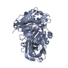 1xn2C 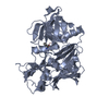 1fknS S: Starting model for refinement C: citing same article ( |
|---|---|
| Similar structure data |


-Links
 PDBj
PDBj

- Assembly
| Deposited unit | 
| ||||||||
|---|---|---|---|---|---|---|---|---|---|
| 1 | 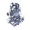
| ||||||||
| 2 | 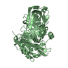
| ||||||||
| 3 | 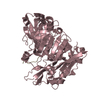
| ||||||||
| 4 | 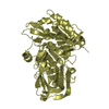
| ||||||||
| Unit cell |
|
-Components
| #1: Protein | Mass: 43312.805 Da / Num. of mol.: 4 / Fragment: Catalytic domain of beta-secretase Source method: isolated from a genetically manipulated source Source: (gene. exp.) Homo sapiens (human) / Gene: BACE1, BACE / Plasmid: PET11A / Species (production host): Escherichia coli / Production host:  #2: Protein/peptide | | Mass: 1652.815 Da / Num. of mol.: 1 / Source method: obtained synthetically #3: Water | ChemComp-HOH / |  Mass: 18.015 Da / Num. of mol.: 744 / Source method: isolated from a natural source / Formula: H2O Mass: 18.015 Da / Num. of mol.: 744 / Source method: isolated from a natural source / Formula: H2OHas protein modification | Y | |
|---|
-Experimental details
-Experiment
| Experiment | Method: X-RAY DIFFRACTION / Number of used crystals: 1 |
|---|
- Sample preparation
| Crystal | Density Matthews: 2.9 Å3/Da / Density % sol: 55.6 % |
|---|---|
| Crystal grow | Temperature: 290 K / Method: vapor diffusion, hanging drop / pH: 6.5 Details: 18% PEG8000, 100mM Cacodylate, pH 6.5, VAPOR DIFFUSION, HANGING DROP, temperature 290K |
-Data collection
| Diffraction | Mean temperature: 100 K |
|---|---|
| Diffraction source | Source: ROTATING ANODE / Type: RIGAKU RU300 / Wavelength: 1.5418 Å |
| Detector | Type: RIGAKU RAXIS IV / Detector: IMAGE PLATE / Date: Jul 8, 2003 / Details: mirrors |
| Radiation | Monochromator: Ni MIRROR + Ni FILTER / Protocol: SINGLE WAVELENGTH / Monochromatic (M) / Laue (L): M / Scattering type: x-ray |
| Radiation wavelength | Wavelength: 1.5418 Å / Relative weight: 1 |
| Reflection | Resolution: 2→50 Å / Num. all: 131154 / Num. obs: 131092 / % possible obs: 99.7 % / Observed criterion σ(I): -3 / Redundancy: 3.8 % / Biso Wilson estimate: 23.5 Å2 / Rmerge(I) obs: 0.079 / Rsym value: 0.079 / Net I/σ(I): 14.5 |
| Reflection shell | Resolution: 2→2.07 Å / Redundancy: 3.5 % / Rmerge(I) obs: 0.384 / Mean I/σ(I) obs: 3.3 / Num. unique all: 12975 / Rsym value: 0.384 / % possible all: 99.2 |
- Processing
| Software |
| ||||||||||||||||
|---|---|---|---|---|---|---|---|---|---|---|---|---|---|---|---|---|---|
| Refinement | Method to determine structure: MOLECULAR REPLACEMENT Starting model: pdb entry 1FKN Resolution: 2→50 Å / Isotropic thermal model: Isotropic / Cross valid method: THROUGHOUT / σ(F): 0 / Stereochemistry target values: Engh & Huber
| ||||||||||||||||
| Displacement parameters | Biso mean: 25.5 Å2 | ||||||||||||||||
| Refinement step | Cycle: LAST / Resolution: 2→50 Å
| ||||||||||||||||
| Refine LS restraints |
|
