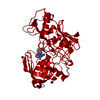
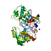
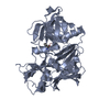
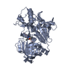
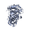
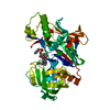
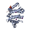
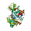
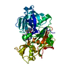
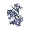
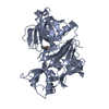
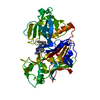
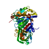
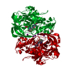
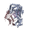
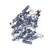
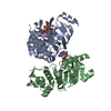
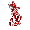
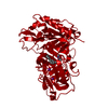
Entry Database : PDB / ID : 2va7Title X-ray crystal structure of beta secretase complexed with compound 27 BETA-SECRETASE 1 . Keywords / / / / / / / / / / / / Function / homology Function Domain/homology Component
/ / / / / / / / / / / / / / / / / / / / / / / / / / / / / / / / / / / / / / / / / / / / / / / / / / / / / / / / / / / / / / / / / / / / / / / / / / / / / Biological species HOMO SAPIENS (human)Method / / / Resolution : 2.2 Å Authors Edwards, P.D. / Albert, J.S. / Sylvester, M. / Aharony, D. / Andisik, D. / Callaghan, O. / Campbell, J.B. / Carr, R.A. / Chessari, G. / Congreve, M. ...Edwards, P.D. / Albert, J.S. / Sylvester, M. / Aharony, D. / Andisik, D. / Callaghan, O. / Campbell, J.B. / Carr, R.A. / Chessari, G. / Congreve, M. / Frederickson, M. / Folmer, R.H.A. / Geschwindner, S. / Koether, G. / Kolmodin, K. / Krumrine, J. / Mauger, R.C. / Murray, C.W. / Olsson, L.L. / Patel, S. / Spear, N. / Tian, G. Journal : J.Med.Chem. / Year : 2007Title : Application of Fragment-Based Lead Generation to the Discovery of Novel, Cyclic Amidine Beta-Secretase Inhibitors with Nanomolar Potency, Cellular Activity, and High Ligand Efficiency.Authors: Edwards, P.D. / Albert, J.S. / Sylvester, M. / Aharony, D. / Andisik, D. / Callaghan, O. / Campbell, J.B. / Carr, R.A. / Chessari, G. / Congreve, M. / Frederickson, M. / Folmer, R.H.A. / ... Authors : Edwards, P.D. / Albert, J.S. / Sylvester, M. / Aharony, D. / Andisik, D. / Callaghan, O. / Campbell, J.B. / Carr, R.A. / Chessari, G. / Congreve, M. / Frederickson, M. / Folmer, R.H.A. / Geschwindner, S. / Koether, G. / Kolmodin, K. / Krumrine, J. / Mauger, R.C. / Murray, C.W. / Olsson, L.L. / Patel, S. / Spear, N. / Tian, G. History Deposition Aug 30, 2007 Deposition site / Processing site Revision 1.0 Nov 13, 2007 Provider / Type Revision 1.1 May 8, 2011 Group Revision 1.2 Jul 13, 2011 Group Revision 1.3 Dec 13, 2023 Group Data collection / Database references ... Data collection / Database references / Other / Refinement description Category chem_comp_atom / chem_comp_bond ... chem_comp_atom / chem_comp_bond / database_2 / pdbx_database_status / pdbx_initial_refinement_model Item / _database_2.pdbx_database_accession / _pdbx_database_status.status_code_sfRevision 1.4 Oct 16, 2024 Group / Structure summaryCategory / pdbx_modification_feature / softwareItem / _software.name
Show all Show less Remark 700 SHEET THE SHEET STRUCTURE OF THIS MOLECULE IS BIFURCATED. IN ORDER TO REPRESENT THIS FEATURE IN ... SHEET THE SHEET STRUCTURE OF THIS MOLECULE IS BIFURCATED. IN ORDER TO REPRESENT THIS FEATURE IN THE SHEET RECORDS BELOW, TWO SHEETS ARE DEFINED.
 Movie
Movie Controller

 Yorodumi
 Open data
 Basic information
 Components
 Keywords
 Function and homology information
 HOMO SAPIENS (human)
 X-RAY DIFFRACTION /
  Authors
 Citation
 Structure visualization
 Downloads & links
 Other downloads
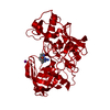


 PDBj
PDBj

 Assembly
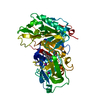


 Mass: 126.904 Da / Num. of mol.: 3 / Source method: obtained synthetically / Formula: I
 Mass: 351.442 Da / Num. of mol.: 1 / Source method: obtained synthetically / Formula: C21H25N3O2
 Mass: 18.015 Da / Num. of mol.: 203 / Source method: isolated from a natural source / Formula: H2O
 Sample preparation
 / Beamline: ID14-3 / Wavelength: 0.931
  Processing