[English] 日本語
Yorodumi Papers
- Database of articles cited by EMDB/PDB/SASBDB data -

+
Search query

Keywords
Structure methods
Author
Journal
IF

-
Structure paper

TitleUncovering druggable hotspots on Pin1 via X-ray crystallographic fragment screening.
Journal, issue, pagesEur. J. Med. Chem., Vol. 299, Page 118048-118048, Year 2025
Publish dateOct 12, 2024 (structure data deposition date)
AuthorsXiao, Q. / Tang, J. / Shu, H. / Wu, T. / Zhang, H. / Wang, W. / Wang, L. / Qin, W.
External linksEur. J. Med. Chem. / PubMed:40803165
MethodsX-ray diffraction
Resolution1.53 - 2.49 Å
Structure data

PDB-9jyo:
Crystal structure of the Pin1 and fragment 2 complex.
Method: X-RAY DIFFRACTION / Resolution: 1.6 Å

PDB-9jyp:
Crystal structure of the PIN1 and fragment 3 complex
Method: X-RAY DIFFRACTION / Resolution: 1.6 Å

PDB-9jyr:
Crystal structure of the PIN1 and fragment 4 complex
Method: X-RAY DIFFRACTION / Resolution: 1.65 Å

PDB-9jyt:
Crystal structure of the PIN1 and fragment 1 complex.
Method: X-RAY DIFFRACTION / Resolution: 1.85 Å

PDB-9jyv:
Crystal structure of the PIN1 and fragment 6 complex.
Method: X-RAY DIFFRACTION / Resolution: 1.8 Å

PDB-9jz2:
Crystal structure of the PIN1 and fragment 7 complex.
Method: X-RAY DIFFRACTION / Resolution: 1.55 Å

PDB-9jz3:
Crystal structure of the PIN1 and fragment 8 complex.
Method: X-RAY DIFFRACTION / Resolution: 1.98 Å

PDB-9jz4:
Crystal structure of the PIN1 and fragment 9 complex.
Method: X-RAY DIFFRACTION / Resolution: 1.82 Å

PDB-9jz6:
Crystal structure of the PIN1 and fragment 11 complex.
Method: X-RAY DIFFRACTION / Resolution: 1.65 Å

PDB-9jzg:
Crystal structure of the PIN1 and fragment 12 complex.
Method: X-RAY DIFFRACTION / Resolution: 1.76 Å

PDB-9jzs:
Crystal structure of the PIN1 and fragment 13 complex.
Method: X-RAY DIFFRACTION / Resolution: 1.91 Å

PDB-9jzu:
Crystal structure of the PIN1 and fragment 14 complex.
Method: X-RAY DIFFRACTION / Resolution: 1.68 Å

PDB-9jzv:
Crystal structure of the PIN1 and fragment 15 complex.
Method: X-RAY DIFFRACTION / Resolution: 1.9 Å

PDB-9ke7:
Crystal structure of the PIN1 and fragment 23 complex.
Method: X-RAY DIFFRACTION / Resolution: 1.68 Å

PDB-9ke9:
Crystal structure of the PIN1 and fragment 22 complex.
Method: X-RAY DIFFRACTION / Resolution: 2.25 Å

PDB-9keb:
Crystal structure of the PIN1 and fragment 24 complex.
Method: X-RAY DIFFRACTION / Resolution: 1.65 Å

PDB-9kec:
Crystal structure of the PIN1 and fragment 25 complex.
Method: X-RAY DIFFRACTION / Resolution: 1.68 Å

PDB-9kel:
Crystal structure of the PIN1 and fragment 26 complex.
Method: X-RAY DIFFRACTION / Resolution: 1.96 Å

PDB-9keq:
Crystal structure of the PIN1 and fragment 27 complex.
Method: X-RAY DIFFRACTION / Resolution: 1.53 Å

PDB-9kes:
Crystal structure of the PIN1 and fragment 28 complex.
Method: X-RAY DIFFRACTION / Resolution: 1.62 Å

PDB-9kew:
Crystal structure of the PIN1 and fragment 29 complex.
Method: X-RAY DIFFRACTION / Resolution: 1.6 Å

PDB-9key:
Crystal structure of the PIN1 and fragment 30 complex.
Method: X-RAY DIFFRACTION / Resolution: 2.49 Å

PDB-9kez:
Crystal structure of the PIN1 and fragment 31 complex.
Method: X-RAY DIFFRACTION / Resolution: 1.95 Å

PDB-9kf0:
Crystal structure of the PIN1 and fragment 32 complex.
Method: X-RAY DIFFRACTION / Resolution: 1.82 Å

PDB-9kfc:
Crystal structure of the PIN1 and fragment 40 complex.
Method: X-RAY DIFFRACTION / Resolution: 1.56 Å

PDB-9kfh:
Crystal structure of the PIN1 and fragment 52 complex
Method: X-RAY DIFFRACTION / Resolution: 1.59 Å

PDB-9kg9:
Crystal structure of the PIN1 and fragment 18 complex.
Method: X-RAY DIFFRACTION / Resolution: 1.55 Å

PDB-9kx7:
Crystal structure of the PIN1 and fragment 34 complex.
Method: X-RAY DIFFRACTION / Resolution: 1.53 Å

PDB-9kx9:
Crystal structure of the PIN1 and fragment 35 complex.
Method: X-RAY DIFFRACTION / Resolution: 1.62 Å

PDB-9kxc:
Crystal structure of the PIN1 and fragment 36 complex.
Method: X-RAY DIFFRACTION / Resolution: 1.53 Å

PDB-9kxd:
Crystal structure of the PIN1 and fragment 37 complex.
Method: X-RAY DIFFRACTION / Resolution: 1.6 Å

PDB-9kxe:
Crystal structure of the PIN1 and fragment 38 complex.
Method: X-RAY DIFFRACTION / Resolution: 1.61 Å

PDB-9kxf:
Crystal structure of the PIN1 and fragment 39 complex.
Method: X-RAY DIFFRACTION / Resolution: 1.71 Å

PDB-9kxg:
Crystal structure of the PIN1 and fragment 41 complex.
Method: X-RAY DIFFRACTION / Resolution: 1.58 Å

PDB-9kxh:
Crystal structure of the PIN1 and fragment 42 complex
Method: X-RAY DIFFRACTION / Resolution: 1.68 Å

PDB-9kxi:
Crystal structure of the PIN1 and fragment 31 complex.
Method: X-RAY DIFFRACTION / Resolution: 1.77 Å

PDB-9kxj:
Crystal structure of the PIN1 and fragment 53 complex
Method: X-RAY DIFFRACTION / Resolution: 1.57 Å

PDB-9kxl:
Crystal structure of the PIN1 and fragment 56 complex.
Method: X-RAY DIFFRACTION / Resolution: 1.7 Å

PDB-9kxm:
Crystal structure of the PIN1 and fragment 57 complex.
Method: X-RAY DIFFRACTION / Resolution: 1.64 Å

PDB-9kxn:
Crystal structure of the PIN1 and fragment 58 complex.
Method: X-RAY DIFFRACTION / Resolution: 1.67 Å

PDB-9kxo:
Crystal structure of the PIN1 and fragment 21 complex.
Method: X-RAY DIFFRACTION / Resolution: 1.75 Å

PDB-9kxp:
Crystal structure of the PIN1 and fragment 33 complex.
Method: X-RAY DIFFRACTION / Resolution: 1.81 Å

PDB-9kxq:
Crystal structure of the PIN1 and fragment 55 complex.
Method: X-RAY DIFFRACTION / Resolution: 1.69 Å

PDB-9v92:
Crystal structure of the Pin1 and Frag61 complex
Method: X-RAY DIFFRACTION / Resolution: 1.97 Å

Chemicals

PDB-1edy:
CRYSTAL STRUCTURE OF RAT ALPHA 1-MACROGLOBULIN RECEPTOR BINDING DOMAIN

ChemComp-PE8:
3,6,9,12,15,18,21-HEPTAOXATRICOSANE-1,23-DIOL

ChemComp-SO4:
SULFATE ION

ChemComp-CL:
Unknown entry

ChemComp-HOH:
WATER

PDB-1edz:
STRUCTURE OF THE NAD-DEPENDENT 5,10-METHYLENETETRAHYDROFOLATE DEHYDROGENASE FROM SACCHAROMYCES CEREVISIAE

ChemComp-P33:
3,6,9,12,15,18-HEXAOXAICOSANE-1,20-DIOL / precipitant*YM

PDB-1ed0:
NMR structural determination of viscotoxin A3 from Viscum album L.

PDB-1edx:
SOLUTION STRUCTURE OF AMINO TERMINUS OF BOVINE RHODOPSIN (RESIDUES 1-40)

PDB-1ed2:
Unknown entry

PDB-1ed3:
CRYSTAL STRUCTURE OF RAT MINOR HISTOCOMPATIBILITY ANTIGEN COMPLEX RT1-AA/MTF-E.

PDB-1ed4:
BOVINE ENDOTHELIAL NITRIC OXIDE SYNTHASE HEME DOMAIN COMPLEXED WITH IPITU (H4B FREE)

PDB-1ed5:
BOVINE ENDOTHELIAL NITRIC OXIDE SYNTHASE HEME DOMAIN COMPLEXED WITH NNA(H4B FREE)

ChemComp-PE4:
2-{2-[2-(2-{2-[2-(2-ETHOXY-ETHOXY)-ETHOXY]-ETHOXY}-ETHOXY)-ETHOXY]-ETHOXY}-ETHANOL / precipitant*YM

PDB-1ed7:
SOLUTION STRUCTURE OF THE CHITIN-BINDING DOMAIN OF BACILLUS CIRCULANS WL-12 CHITINASE A1

PDB-1ed8:
STRUCTURE OF E. COLI ALKALINE PHOSPHATASE INHIBITED BY THE INORGANIC PHOSPHATE AT 1.75A RESOLUTION

PDB-1at6:
HEN EGG WHITE LYSOZYME WITH A ISOASPARTATE RESIDUE

PDB-1ed9:
STRUCTURE OF E. COLI ALKALINE PHOSPHATASE WITHOUT THE INORGANIC PHOSPHATE AT 1.75A RESOLUTION

PDB-1eea:
Acetylcholinesterase

PDB-1ee7:
NMR STRUCTURE OF THE PEPTAIBOL CHRYSOSPERMIN C BOUND TO DPC MICELLES

PDB-1bdv:
ARC FV10 COCRYSTAL

PDB-1ee8:
CRYSTAL STRUCTURE OF MUTM (FPG) PROTEIN FROM THERMUS THERMOPHILUS HB8

PDB-1ee6:
CRYSTAL STRUCTURE OF PECTATE LYASE FROM BACILLUS SP. STRAIN KSM-P15.

PDB-1efa:
CRYSTAL STRUCTURE OF THE LAC REPRESSOR DIMER BOUND TO OPERATOR AND THE ANTI-INDUCER ONPF

PDB-1efb:
Unknown entry

PDB-1efc:
INTACT ELONGATION FACTOR FROM E.COLI

PDB-1efd:
PERIPLASMIC FERRIC SIDEROPHORE BINDING PROTEIN FHUD COMPLEXED WITH GALLICHROME

PDB-1efe:
AN ACTIVE MINI-PROINSULIN, M2PI

ChemComp-DBJ:
2,3-dihydro-1,4-benzodioxin-5-ylmethanol

PDB-1eff:
Unknown entry

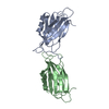
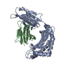
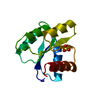
PDB-1aya:
CRYSTAL STRUCTURES OF PEPTIDE COMPLEXES OF THE AMINO-TERMINAL SH2 DOMAIN OF THE SYP TYROSINE PHOSPHATASE

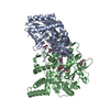
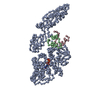
PDB-1efg:
THE CRYSTAL STRUCTURE OF ELONGATION FACTOR G COMPLEXED WITH GDP, AT 2.7 ANGSTROMS RESOLUTION

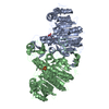
PDB-1efp:
ELECTRON TRANSFER FLAVOPROTEIN (ETF) FROM PARACOCCUS DENITRIFICANS

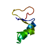
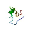
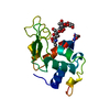
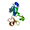
PDB-1ehc:
STRUCTURE OF SIGNAL TRANSDUCTION PROTEIN CHEY

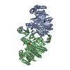
PDB-1ehd:
CRYSTAL STRUCTURE OF URTICA DIOICA AGGLUTININ ISOLECTIN VI

PDB-1ehe:
CRYSTAL STRUCTURES OF CYTOCHROME P450NOR AND ITS MUTANTS (SER286 VAL, THR) IN THE FERRIC RESTING STATE AT CRYOGENIC TEMPERATURE: A COMPARATIVE ANALYSIS WITH MONOOXYGENASE CYTOCHROME P450S

ChemComp-53E:
2,3-dihydro-1,4-benzodioxine-5-carboxylic acid

PDB-1ehf:
CRYSTAL STRUCTURES OF CYTOCHROME P450NOR AND ITS MUTANTS (SER286 VAL, THR) IN THE FERRIC RESTING STATE AT CRYOGENIC TEMPERATURE: A COMPARATIVE ANALYSIS WITH MONOOXYGENASE CYTOCHROME P450S

PDB-1ehg:
CRYSTAL STRUCTURES OF CYTOCHROME P450NOR AND ITS MUTANTS (SER286 VAL, THR) IN THE FERRIC RESTING STATE AT CRYOGENIC TEMPERATURE: A COMPARATIVE ANALYSIS WITH MONOOXYGENASE CYTOCHROME P450S

ChemComp-M4T:
~{N}-pyridin-3-ylethanamide

PDB-1eho:
Unknown entry

PDB-1ehy:
X-ray structure of the epoxide hydrolase from agrobacterium radiobacter ad1

PDB-1evl:
CRYSTAL STRUCTURE OF A TRUNCATED FORM OF THREONYL-TRNA SYNTHETASE WITH A THREONYL ADENYLATE ANALOG

PDB-1ehq:
Unknown entry

PDB-1erz:
CRYSTAL STRUCTURE OF N-CARBAMYL-D-AMINO ACID AMIDOHYDROLASE WITH A NOVEL CATALYTIC FRAMEWORK COMMON TO AMIDOHYDROLASES

PDB-1ehr:
Unknown entry

ChemComp-UUY:
2-[methyl(pyridin-2-yl)amino]ethan-1-ol

PDB-1ehb:
CRYSTAL STRUCTURE OF RECOMBINANT TRYPSIN-SOLUBILIZED FRAGMENT OF CYTOCHROME B5


PDB Unreleased entry

PDB-1ehp: CRYSTAL STRUCTURE OF A TERNARY COMPLEX OF CAP, DNA AND C-TERMINAL DOMAIN OF ALPHA SUBUNIT OF RNA POLYMERASE FRO E. COLI.


ChemComp, No image

ChemComp-QCF:
Unknown entry

Source
  • homo sapiens (human)
  • escherichia coli bl21(de3) (bacteria)
KeywordsISOMERASE / cis-trans isomerase / Inhibitor

+
About Yorodumi Papers

-
News

-
Feb 9, 2022. New format data for meta-information of EMDB entries

New format data for meta-information of EMDB entries

  • Version 3 of the EMDB header file is now the official format.
  • The previous official version 1.9 will be removed from the archive.

Related info.:EMDB header

External links:wwPDB to switch to version 3 of the EMDB data model

-
Aug 12, 2020. Covid-19 info

Covid-19 info

URL: https://pdbj.org/emnavi/covid19.php

New page: Covid-19 featured information page in EM Navigator.

Related info.:Covid-19 info / Mar 5, 2020. Novel coronavirus structure data

+
Mar 5, 2020. Novel coronavirus structure data

Novel coronavirus structure data

Related info.:Yorodumi Speices / Aug 12, 2020. Covid-19 info

External links:COVID-19 featured content - PDBj / Molecule of the Month (242):Coronavirus Proteases

+
Jan 31, 2019. EMDB accession codes are about to change! (news from PDBe EMDB page)

EMDB accession codes are about to change! (news from PDBe EMDB page)

  • The allocation of 4 digits for EMDB accession codes will soon come to an end. Whilst these codes will remain in use, new EMDB accession codes will include an additional digit and will expand incrementally as the available range of codes is exhausted. The current 4-digit format prefixed with “EMD-” (i.e. EMD-XXXX) will advance to a 5-digit format (i.e. EMD-XXXXX), and so on. It is currently estimated that the 4-digit codes will be depleted around Spring 2019, at which point the 5-digit format will come into force.
  • The EM Navigator/Yorodumi systems omit the EMD- prefix.

Related info.:Q: What is EMD? / ID/Accession-code notation in Yorodumi/EM Navigator

External links:EMDB Accession Codes are Changing Soon! / Contact to PDBj

+
Jul 12, 2017. Major update of PDB

Major update of PDB

  • wwPDB released updated PDB data conforming to the new PDBx/mmCIF dictionary.
  • This is a major update changing the version number from 4 to 5, and with Remediation, in which all the entries are updated.
  • In this update, many items about electron microscopy experimental information are reorganized (e.g. em_software).
  • Now, EM Navigator and Yorodumi are based on the updated data.

External links:wwPDB Remediation / Enriched Model Files Conforming to OneDep Data Standards Now Available in the PDB FTP Archive

-
Yorodumi Papers

Database of articles cited by EMDB/PDB/SASBDB data

  • Database of articles cited by EMDB, PDB, and SASBDB entries
  • Using PubMed data

Related info.:EMDB / PDB / SASBDB / Yorodumi / EMN Papers / Changes in new EM Navigator and Yorodumi

Read more